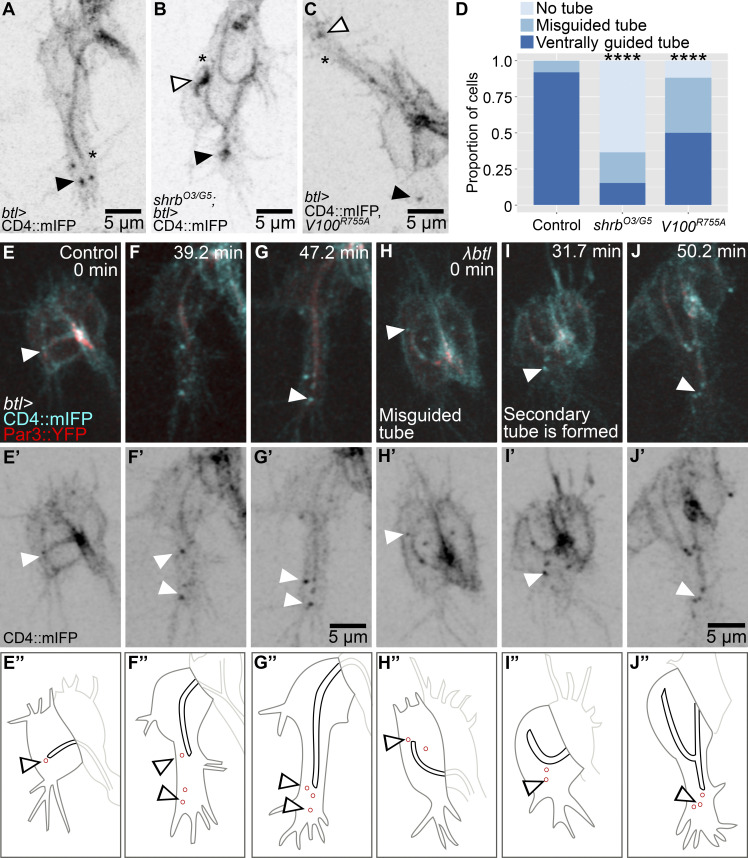
Figure 1.
Distribution and role of late endosomes in subcellular tube guidance. (A–D) Terminal cells expressing CD4::mIFP under btl-gal4; see also Video 1. (A) Control. (B) Cell mutant for shrb. (C) Cell expressing Vha100R755A (V100R755A), a dominant negative construct that blocks vATPase function. (D) Proportion of cells with the indicated phenotypes. Number of cells analyzed: control, n = 37; shrbO3/G5, n = 33; Vha100R755A, n = 42. Significance was assayed with a χ2 test; ****, P < 0.0001. (E–J) Terminal cells expressing CD4::mIFP and Par3::YFP under btl-gal4. (E’–J’) CD4::mIFP. (E’’–J’’) Manual tracings of the contours of the terminal cells (dark gray), subcellular tube (black), endosomes (red), and the adjacent dorsal branch cells (light gray). (E–G) Control. (H–J) Cells expressing a constitutively active FGFR, λbtl. Asterisks in A–C mark the tips of the subcellular tubes, and black and white arrowheads point to CD4 vesicles. Ventral is down.

