Figure 7.
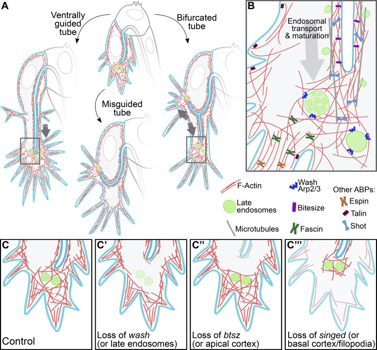
Graphic summary of the role of late endosomes in subcellular tube guidance. (A) Control cells. Late endosomes situated at the tip of the cell allow the organization of an actin network that extends from the tube to the growing tip of the cell. Control cells with misguided tubes result from the nucleation of actin surrounding late endosomes not located at the tip. In cells with tube bifurcations, late endosomes form two actin nucleation centers that stabilize each subcellular branch. (B) Zoom-in to the tip of the cell (regions marked with rectangles in A). Distinct actin-binding proteins (ABPs) at different subcellular compartments coordinate proper tube formation, with Wash acting in late endosomes to bridge actin pools at the apical (via Btsz) and basal (via Talin and the filopodial organizers Fascin and Espin) cortices. Also shown are microtubules that surround the subcellular tube and that are anchored to the actin meshwork at the tip via Shot. (C–C’’’) Effects of loss of actin organizers at distinct subcellular compartments. (C) Control. (C’) Upon loss of wash or in conditions with aberrant endosomal maturation, F-actin fails to form around late endosomes. (C’’) In the absence of btsz or other actin organizers at the apical cortex, F-actin fails to cross-link to the actin meshwork at the tip. (C’’’) Lack of actin regulators at the basal cortex prevents the connection of the actin meshwork at the tip to the basal plasma membrane. All these conditions decouple the growth of the tube from that of the tip of the cell.

