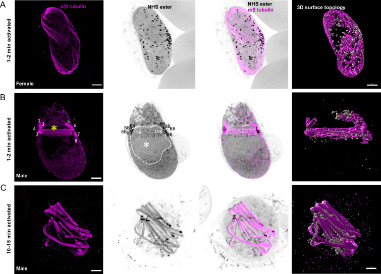
Fig 2. Mitotic structures and axonemes are highlighted by U-ExM during P. falciparum gametogenesis.
A-C. Representative full projections of activated P. falciparum gametocytes. α/β-tubulin: magenta; amine reactive groups / NHS-ester: shades of grey. Column 4 shows the 3D surface topology reconstruction of α/β-tubulin and NHS-ester. A. A 1–2 min activated macrogametocyte shows degrading of the subpellicular microtubules and NHS-ester dense osmiophilic bodies. B. A 1–2 min activated microgametocyte shows the NHS-ester dense basal bodies (BB) giving rise to axonemes (1 to 8 visible in S1 Movie). The mitotic spindle (yellow asterisk) is highlighted by α/β-tubulin staining and at each extremity the intranuclear bodies (INB) show an NHS-ester dense staining. NHS-ester positive kinetochores are present along the spindle. The white dotted line highlights the nuclear periphery. At this stage, the subpellicular microtubules are completely lost. C. A 10–15 min activated microgametocyte displays a round shaped and full length axonemes. Scale bars = 5 μm.

