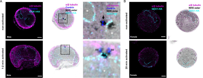
Fig 3. The MTOC coordinating axoneme formation and mitosis shows a bipartite structure across the nuclear membrane during P. berghei microgametogenesis.
A and B. Localisation of the nuclear membrane protein GEX1-HA in P. berghei gametocytes. α/β-tubulin: magenta; amine reactive groups/NHS-ester: shades of grey; centrin: blue; GEX1-HA: cyan. A. Non-activated and activated microgametocytes show a lobulated nuclear architecture, as seen by GEX1-HA signal and tubulin negative areas. Column 3 represents a close up of the boxed areas. The nuclear contour is distinguished by a light NHS-ester staining, which matches GEX1-HA staining. Nucleus: white asterisk; amorphous MTOC: black arrow (centrin-positive); basal bodies: BB (centrin-positive); intranuclear body: INB (centrin-negative). B. Non-activated and activated macrogametocytes. Macrogametocytes display a crescent-shaped nucleus, very low levels of α/β-tubulin, and NHS-ester dense osmiophilic bodies. Scale bars = 5 μm.

