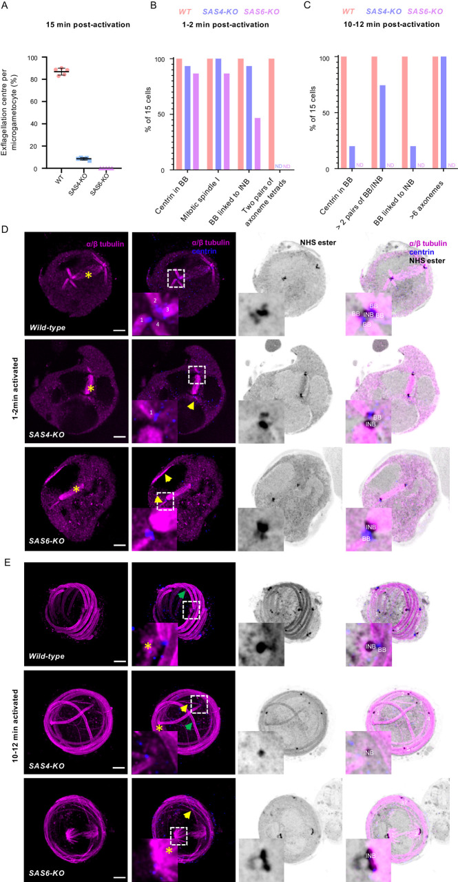
Fig 8. Characterisation of SAS4-KO and SAS6-KO lines highlights different requirements of both proteins for the integrity of the basal body.
A. Deletions of sas4 or sas6 lead to a profound defect in exflagellation (error bars show standard deviation from the mean). B-C. Quantification of the phenotypes shown in D and E in 15 individual cells for each line. BB = basal body; INB = intranuclear body. ND = Not Detected. D-E. Representative full projections of (D) 1–2 min activated and (E) 10–12 min activated microgametocytes; wild-type (1st row), SAS4-KO (2nd row) and SAS6-KO (3rd row). α/β-tubulin: magenta; amine reactive groups/NHS-ester: shades of grey; centrin: blue. Boxed areas correspond to close-ups. Yellow star = mitotic spindle; yellow arrow = non-bundled microtubules; green arrow = bundled microtubules; INB = intranuclear body; BB = basal body. Scale bar = 5 μm.

