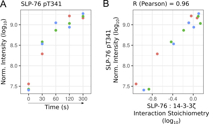
Figure S3.
Phosphorylation dynamics of the threonine residue found at position 341 of SLP-76. (A) Plot showing the log10-transformed intensity for the phospho-site corresponding to the threonine residue found at position 341 of SLP-76 (SLP-76-pT341) over the course of TCR stimulation. Phosphorylated peptide intensity was quantified using AP-MS samples of SLP-76OST human CD4+ T cells. Data corresponding to each of the three biological replicates are specified by dots of a distinct color. Norm., normalized. (B) Plot showing the correlation between SLP-76-pT341 intensity and the stoichiometry of the interaction between SLP-76 and its high-confidence prey 14-3-3ζ, both quantified from AP-MS data. Our dataset did not permit identification of additional phosphorylated peptides with high confidence in the SLP-76 bait and the other baits.

