
FIG 2.
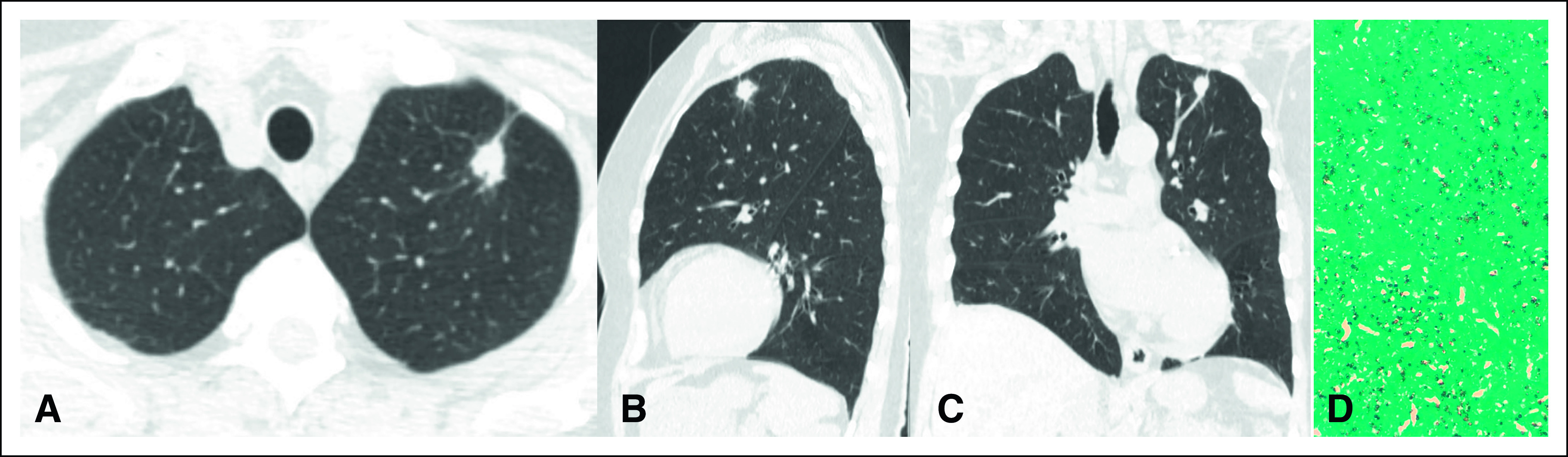
(A) Axial computed tomography image shows a spiculated pulmonary nodule classified as Lung-RADS 4Bx. CT images with (B) coronal and (C) sagittal reconstruction demonstrate the same findings. (D) CT-guided biopsy specimens contained predominantly noncaseating granulomas and intracellular and extracellular fungal elements compatible with histoplasmosis (Grocott, ×400). CT, computed tomography.