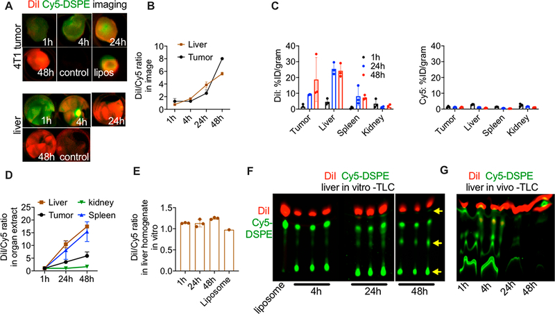
Figure 6.
FPLs are less stable than ICLs and are eliminated from tumor and organs in vivo. (A) Mice were injected with DiI/Cy5-DSPE-labeled liposomes, and tumors (upper panel) and livers (lower panel) were excised and imaged at different time points. Representative images of merged DiI and Cy5 fluorescence are shown (n = 2 mice, repeated three times). Liposomes were dotted on a membrane and scanned together with tumors (image of the liposomal dot is shown in upper panel). (B) The DiI/Cy5 fluorescence ratio in tumor and liver images shows an increase over time (n = 2 mice per time point). (C) Tumors and major organs were homogenized and the lipids were extracted with organic solvent as described in the Methods. DiI (left graph) and Cy5 (right graph) show major differences in biodistribution. (D) The DiI/Cy5 fluorescence ratio in tumor and organ extracts shows an increase over time. (E) Liver homogenates were spiked with DiI/Cy5-DSPE liposomes and incubated for different times. There was no increase in DiI/Cy5 fluorescence ratio over time. (F) Thin layer chromatography analysis of fluorescence after lipid extraction from liver homogenates in (E). The arrow points to degradation of Cy5-DSPE. (G) TLC analysis of liver extracts at different times after injection of DiI/Cy5-DSPE-labeled liposomes shows a decrease in the levels and degradation of Cy5-DSPE but not DiI.

