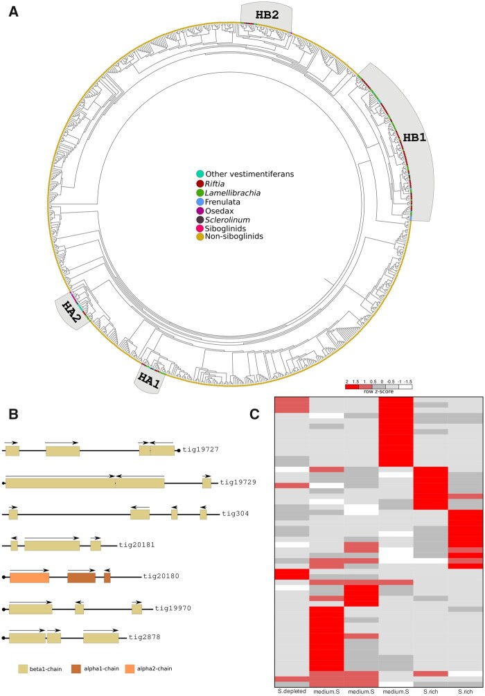
Fig. 3.
Expanded hemoglobin complement in Riftia pachyptila. (A) Midpoint rooted phylogeny of 693 Riftia, annelid and metazoan hemoglobin genes, using Belato et al. (2019) as backbone. Colored circles correspond to different annelid taxa and metazoans. (B) Seven genomic hemoglobin clusters are present in the Riftia genome. Arrows indicate the direction of transcription. Scaffolds with a circle on the end indicate the presence of Hb genes in the terminal end of the genomic segment. Only the longest gene models are shown. Colors represent the different hemoglobin chains. (C) Heat map expression of hemoglobins in the trophosome under three experimental conditions (obtained from Hinzke et al. [2019]): medium sulfide (medium.S), sulfide rich (S.rich), and sulfide depleted (S.depleted).

