. 2022 Jan 4;11:e74993. doi: 10.7554/eLife.74993

Figure 2. ARMC2-3xTAG is highly enriched in regenerating flagella.

(A) TIRF imaging of ARMC2-3xTAG in the pf27 background (a, b) and the pf14 armc2 double mutant background (c, d) in full-length (a, c) and in regenerating flagella (b, d). Bars = 2 s 2 µm. The flagellar tips and bases are indicated. (B) Bar graph showing the average frequencies (events/min/flagellum) of anterograde and retrograde transport of ARMC2-3xTAG in full-length and regenerating flagella of the pf27 ARMC2-3XTAG and the pf14 armc2 ARMC2-3xTAG strain. The standard deviation and the number of flagella analyzed are indicated. (C) Kymograms of ARMC2-3xTAG in late regenerating pf27 flagella. The white brackets in a–c mark the dwell time of individual ARMC2-3xTAG particles between arrival at the tip by anterograde IFT and the onset of diffusion (white arrowheads). Green arrowheads in a–c, anterograde transport of ARMC2-3xTAG, red open arrow in d, retrograde IFT of ARMC2-3xTAG; red arrowheads in e, stepwise bleaching of ARMC2-3xTAG indicating for the presence of two copies. In c, a single step bleaching event is marked by a dashed circle. Bars = 2 s and 2 µm. (D) DIC images of full-length and regenerating flagella of the pf27 ARMC2-3xTAG strain. Regenerating flagella were harvested ~22 min after deflagellation by a pH shock. Bar = 10 µm. (E) Western blot analysis of the full-length and regenerating flagella shown in C with the antibodies indicated. On the left side, an equal number of flagella were loaded and on the right side, approximately equal loading of protein was attempted. (F) Western blot comparing the presence of ARMC2-3xTAG in the pf27 ARMC2-3xTAG and the pf14 armc2 ARMC2-3xTAG strain. Antibodies to the cell body protein nucleic acid binding protein 1 (NAB1) were used as a loading control.

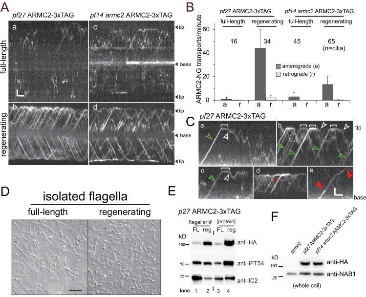
Figure 2.

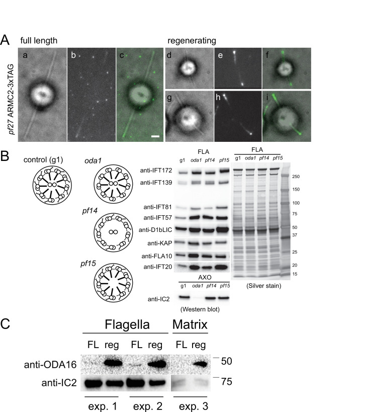
Figure 2—figure supplement 1. ARMC2-3xTAG accumulates at the tip of growing flagella.

Figure 2—figure supplement 1.

(A) Bright field (a, d, g), TIRF (b, e, h), and merged images (c, f, i) of ARMC2-3xTAG in full-length (a–c) and regenerating (d–i) flagella of the pf27 ARMC2-3xTAG strain. Bar = 2 µm. (B) Schematic presentation, Western blot analysis and silver-stained gel of isolated flagella from control (g1), and the oda1, pf14, and pf15 mutants. Note accumulation of intraflagellar transport (IFT) proteins in the flagella these motility mutants. (C) Comparison of ODA16 levels in full-length (FL) and regenerating (reg) wild-type flagella from two independent biological replicates (experiments 1 and 2); from a third experiment (experiment 3) only the matrix fraction was available for analysis. For experiments 1 and 2, approximately equal number of flagella were loaded as apparent from the stronger IC2 bands in the FL samples.
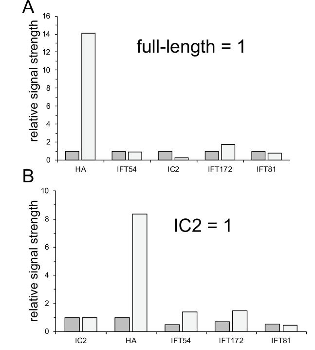
Figure 2—figure supplement 2. ARMC2-3xAG is enriched in growing flagella.

Figure 2—figure supplement 2.

(A, B) Quantification of the Western blot shown in Figure 2E. (A) Quantification of lanes 1 and 2, in which equal number of flagella were loaded. To normalize, the signal obtained with the full-length flagella sample was set to one for each antibody. (B) Quantification of lanes 3 and 4, in which we attempted to load equal amounts of protein. The signals were normalized for the anti-IC2 signal, which was set to one for the full-length and regenerating flagellar sample. The analysis is based on N = 1 Western blots.
Figure 2—video 1. Anterograde intraflagellar transport (IFT) of ARMC2-3xTAG.
Download video file (405.2KB, mp4)
Video and corresponding kymogram showing transport of ARMC2-3xTAG by anterograde IFT to the flagellar tip, dwelling at the tip, and return to the ciliary base by diffusion. The video was recorded at 10 fps and the timer counts seconds. The video is related to Figure 2C.