. 2022 Jan 4;11:e74993. doi: 10.7554/eLife.74993

Figure 5. ARMC2-3xTAG transport is upregulated in short flagella.

(A) Gallery of brightfield (BF) and TIRF still images and the corresponding kymograms of long-short p27 ARMC2−3xTAG cells. No BF image was recorded for the cell shown in a. The length of the long (L) and short (S) flagella is indicated (in µm in a–d). Bars = 2 s and 2 µm. (B) Plot of the ARMC2-3xTAG transport frequency (events/min/flagellum) in the short (squares) and the long (triangles) flagella against the length difference between the two flagella. Trendlines, solid for the long and dashed for the short flagella, were added in Excel. (C) As B, but for cells treated with cycloheximide prior and during the experiment.

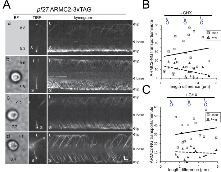
Figure 5.

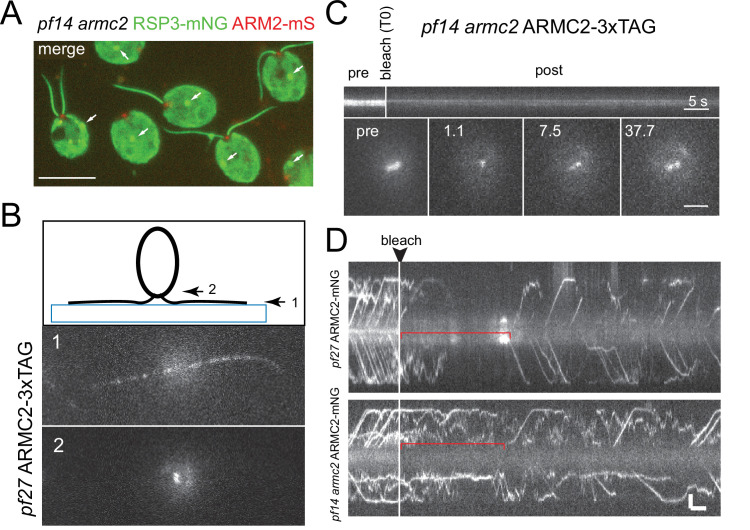
Figure 5—figure supplement 1. ARMC2-FP forms a pool near the basal bodies.

Figure 5—figure supplement 1.

(A) Two-color epifluorescence image of the pf14 armc2 RSP3-NG ARMC2-mS strain. Note accumulation of ARMC2-mS (red) at the flagellar base. The bright signal of the cell body likely results from unspecific binding of the antibody and chlorophyll autofluorescence. The arrows mark the unspecific signal of the eyespot apparatus (arrows). Bar = 10 µm. (B) Focal series using flat-angle ‘TIRF’ illumination showing the flagella level (1) and the two ARMC2-3xTAG signals at the basal body level (2). The diagram illustrates the focal levels of the two images. (C) FRAP experiment using a focused laser beam to bleach one of the two ARMC2-3xTAG signals at the flagella base. Note slow and partial recovery of the signal. Statistical analysis was not performed as only three cells were analyzed. Bars = 5 s and 2 µm. (D) FRAP experiments to determine the dwell time of ARMC2-3xTAG in the basal body-associated pool. Regenerating flagella of the pf27 ARMC2-3xTAG and the pf14 armc2 ARMC2-3xTAG strains were analyzed. As in D, we used a focused laser beam to bleach the entire pool at the flagellar base and then analyzed the length of the gap before IFT of ARMC2-3xTAG resumed.
Figure 5—video 1. ARMC2-3xTAG transport in long-short cells.
Download video file (1.8MB, mp4)
Bright-field and TIRF video and the corresponding kymogram of a long-short pf27 ARMC2-3xTAG cell. Cells were sheared to generate long-zero cells and allowed to regrow missing flagella. The video was recorded at 10 fps and the timer counts seconds. The video is related to Figure 5.
Figure 5—video 2. ARMC2-3xTAG transport in long-short cells.
Download video file (4.6MB, mp4)
Bright-field and TIRF video and the corresponding kymogram of a long-short pf27 ARMC2-3xTAG cell. Cells were treated for 1 hr in CHX, sheared to generate long-zero cells and allowed to regrow missing flagella in the presence of CHX. The video was recorded at 10 fps and the timer counts seconds. Note that the bright-field video loops repeatedly. The video is related to Figure 5