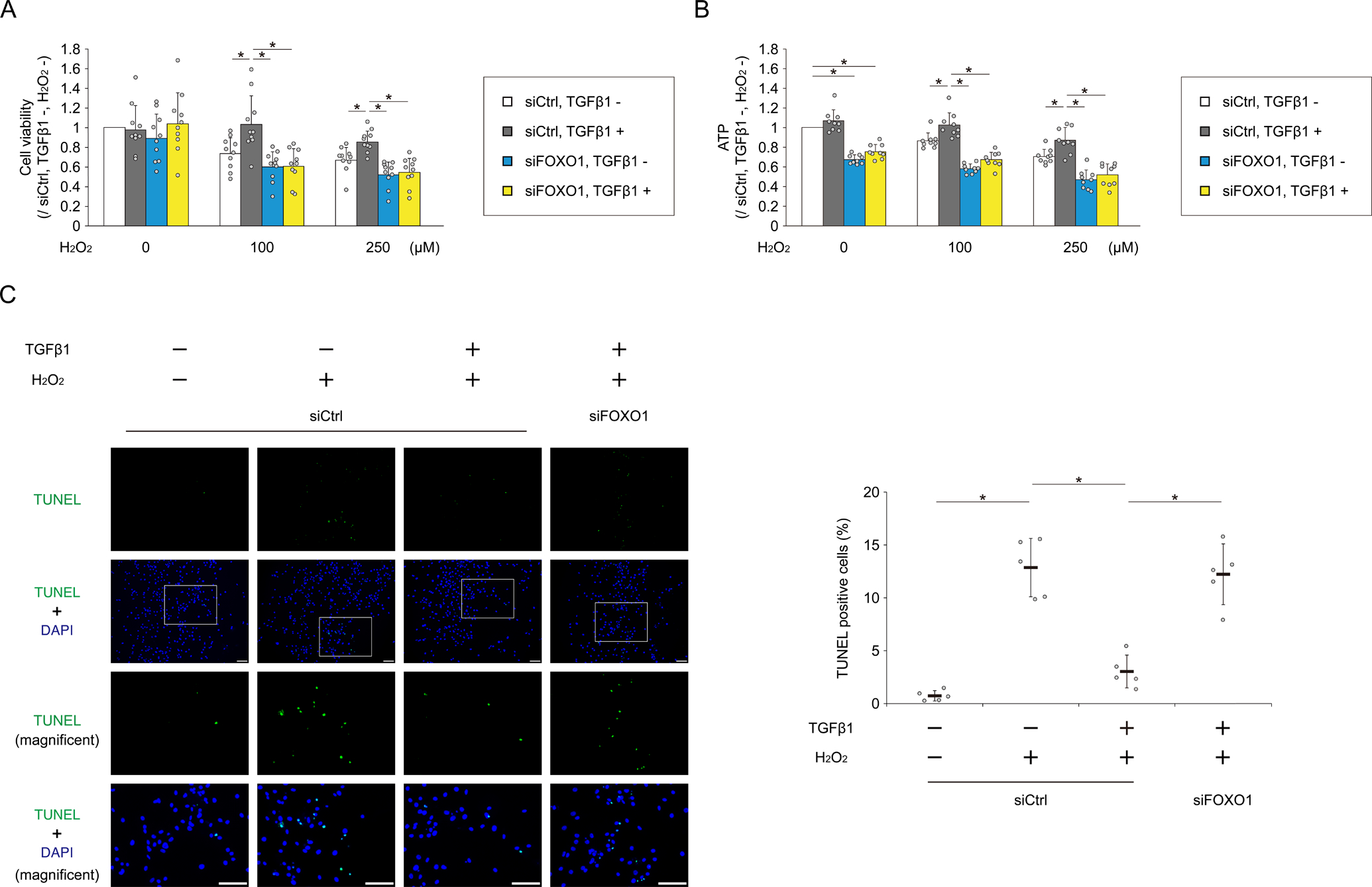
Fig. 6. TGFβ1 protects chondrocytes against oxidative stress–induced cell death via FOXO1.

Human chondrocytes were transfected with siCtrl or siFOXO1. After incubation with or without TGFβ1 for 3 hours, H2O2 was added to each well to a final concentration of 0, 100, or 250 μM for 48 hours. (A) Cell viability was analyzed by MTT assay; n = 10 independent experiments. (B) ATP production was analyzed using the CellTiter-Glo assay; n = 9 independent experiments. Data are expressed as the percent absorbance (A) or luminescence (B) of cells in each group relative to cells transfected with siCtrl, and not treated with TGFβ1 and H2O2. Human chondrocytes were transfected with siCtrl or siFOXO1. After incubation with or without TGFβ1 for 3 hours, H2O2 (0 or 250 μM) was added to each well. After 48 hours, cell apoptosis was analyzed by TUNEL assay (C). Higher-magnification views are corresponding to the white boxed areas in each group. Graph shows the percentage of TUNEL positive cells; n = 5 independent experiments. Data are presented as dots showing individual values and as means ± S.D. Statistical analysis was performed using one-way repeated measures ANOVA with the Tukey–Kramer post hoc test. *P < 0.05.
