
Figure 1.
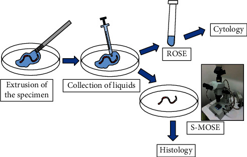
Specimen processing with an endoscopic ultrasound-guided fine-needle biopsy. After each pass, the specimen was extruded from the needle onto a Petri dish with saline. The liquid components around the specimen were aspirated with a syringe and submitted for rapid on-site cytopathological evaluation (ROSE) and cytology. The remaining solid specimens were immediately evaluated under a stereomicroscope and then submitted for histological examination.