
Figure 3.
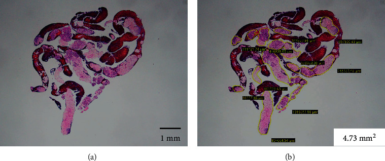
Evaluation of the tissue area using imaging software. (a) Hematoxylin and eosin staining of a gross specimen obtained with endoscopic ultrasound-guided fine-needle biopsy, viewed in a low-power field. (b) Measuring the area of the tissue specimen, excluding the blood clots, using imaging software (CellSens).