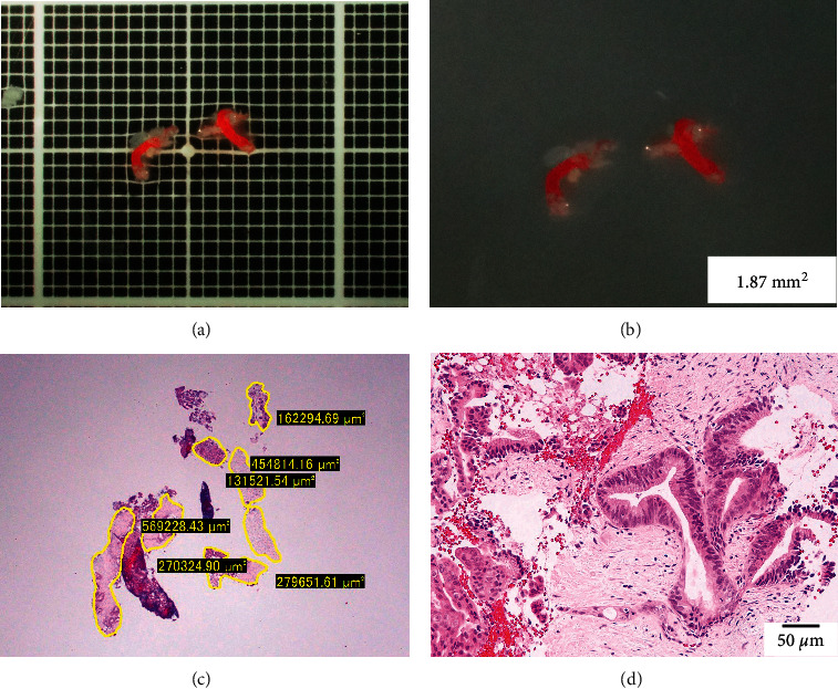
Figure 5.

An example where stereomicroscopic observation was considered useful in determining the presence of core tissue. (a, b) A specimen obtained from a mass in the pancreatic body, observed under a stereomicroscope with (a) and without (b) a black scale. The specimen is relatively small, which is difficult to macroscopically evaluate in detail, but under a stereomicroscope, white core tissue is observed in addition to red blood clots. (c) Measurement using imaging software (CellSens) shows that 1.87 mm2 of tissue was collected. (d) Photomicrograph showing a component of atypical cells with enlarged nuclei in the fibrous stroma, consistent with ductal carcinoma of the pancreas.
