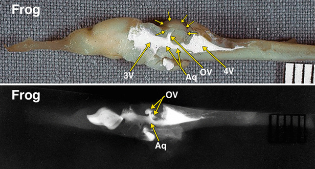
Fig. 2.

Dissected brain of a frog with radiopaque contrast filled cerebral ventricles. The midsagittal dissection (Fig. 2) demonstrates a prominent optic lobe, outlined by multiple small arrows, and includes only a small central segment of the optic ventricles (OV) extending superiorly from the aqueduct (Aq). The remainder of the more laterally positioned optic ventricles is visualized in the radiograph. The lateral radiograph of the entire dissected brain demonstrates both optic ventricles (OV) and the aqueduct (Aq). The constricted communication between the dorsolateral segment of the optic ventricles and medial segment of the opticventricle (aqueduct) is likely the result of compressed from below by the torus semicircularis [28]. Additional abbreviations: 3 V, 3d ventricle; 4 V, 4th ventricle. The distance between 2 black lines on the ruler in the specimen photograph is 1 mm
