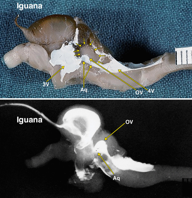
Fig. 3.

Dissected brain of an iguana with radiopaque contrast filled cerebral ventricles. The midsagittal dissection (Fig. 3) demonstrates a relatively large optic lobe, outlined by multiple small arrows, that includes an optic ventricle (OV). The midbrain ventricle (Aq) and optic ventricles form a large continuous cavity. The lateral radiograph (Fig. 3B) of the entire dissected brain demonstrates the mostly superimposed optic ventricles (OV) and the aqueduct (Aq). Additional abbreviations: 3 V, 3d ventricle; 4 V, 4th ventricle. The distance between 2 black lines on the ruler in the specimen photograph is 1 mm
