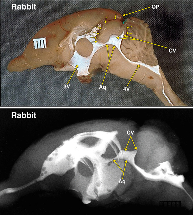
Fig. 4.

Dissected brain of a rabbit with radiopaque contrast filled cerebral ventricles. The midsagittal dissection (Fig. 4) demonstrates large midbrain colliculi outlined by multiple small arrows. A small cleft indicated by a small green arrow likely indicates the separate superior and inferior colliculi of the quadrigeminal plate. The aqueduct (Aq) and collicular ventricle (CV) form a large continuous cavity. Of note is the separate round extension of the collicular ventricle into what is likely the superior, and the pointed extension into the inferior colliculi. Also of note is the presence of a small neocortical occipital lobe and occipital pole (OP). The collicular ventricle features are also demonstrated in the lateral radiograph. Additional abbreviations: 3 V, 3d ventricle; 4 V, 4th ventricle. The distance between 2 black lines on the ruler in the specimen photograph is 1 mm
