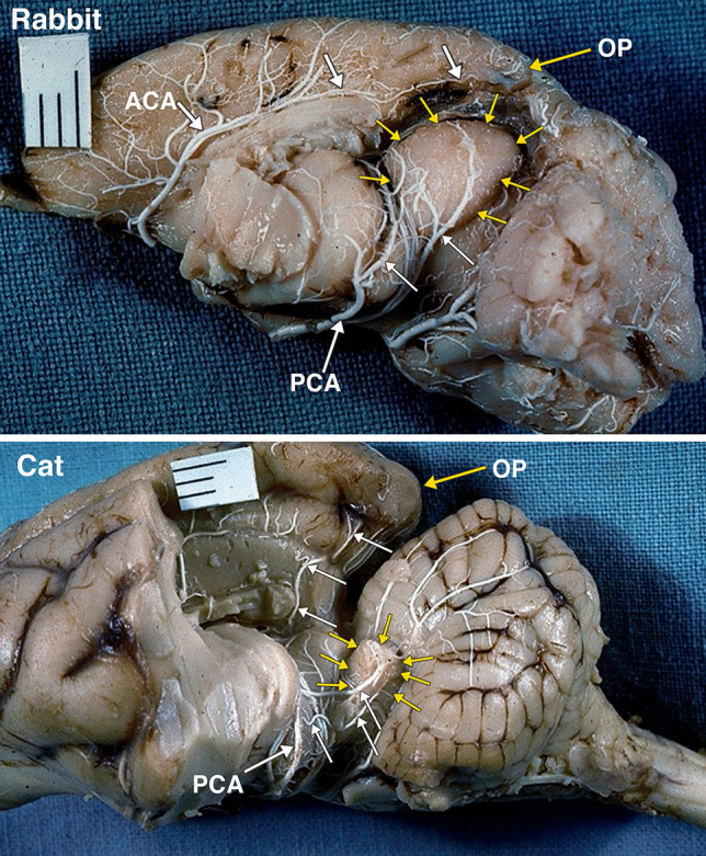
Fig. 8.

A postero-lateral view of an arterially injected and dissected left hemisphere of the brain of an adult rabbit. Identified are the large midbrain colliculi, outlined by yellow arrows. The colliculi are supplied by branches (white arrows) of the posterior cerebral artery (PCA). Of note is the small size of the occipital lobe and pole (OP) supplied by branches (white arrows) of the anterior cerebral artery (ACA). A postero-lateral view of an arterially injected and dissected left hemisphere of the brain of an adult cat. When compared to the rabbit (Fig. 8), the midbrain colliculi, outlined by yellow arrows are much reduced in size. The significant reduction in the relative size of colliculi is accompanied by significant increase in size of the occipital lobe and occipital pole (OP). The branches (white arrows) of the posterior cerebral artery (PCA) supply the colliculi, and also a large region of the occipital lobe. It was not possible to separately identify the superior and inferior colliculi without disrupting the arterial supply embedded in the arachnoid layer
