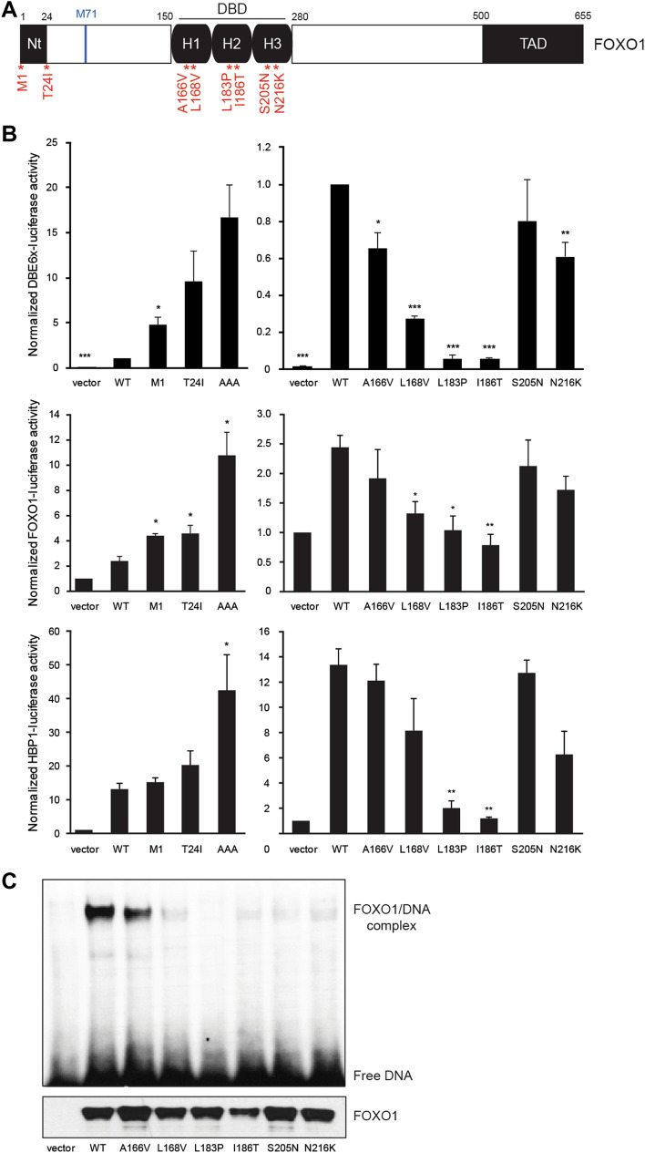
Figure 1.
Transcriptional activity and DNA binding of FOXO1 Nt and DBD mutants. (A) Schematic structure of the human FOXO1 protein and the studied point mutations. (B) HEK293T cells were co-transfected with wild-type or mutated pCMV-FOXO1, luciferase reporter (DBE6x, FOXO1 promoter, HBP1 promoter) and the pEF-β-galactosidase vector as internal control. After 24 h of transfection, cells were lysed to measure the luminescence and the β-galactosidase activity. Average of 3 independent experiments is shown with standard error of the mean. Statistical analysis was calculated according to bilateral Student t-test (*, p < 0.05; **, p < 0.01; ***, p < 0.001). (C) Electrophoretic mobility shift assays (EMSA) were performed using nuclear extracts from HEK293T cells transfected with wild-type or mutated pCMV-FOXO1. A 32P-labeled oligonucleotide probe containing the FOXO consensus DNA-binding sequence (GTAAACA) was added to each nuclear extract. In addition, expression of FOXO1 was analyzed by western blot with an anti-FOXO1 antibody. Original unprocessed images are available in Supplementary Fig. S2.

