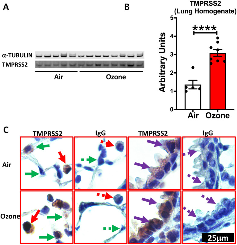
Figure 1.

TMPRSS2 protein expression is upregulated in the lungs of ozone -exposed mice. Western blot representative gel image (A) showing bands for TMPRSS2 protein and alpha-tubulin loading control, and band intensity analyses (B, Bar Graph) on the whole lung homogenate from air- and ozone-exposed mice (n = 6–8). (C) Immunohistochemical staining for TMPRSS2 in macrophages (solid red arrow), alveolar epithelial cells (solid green arrow), and bronchiolar epithelial cells (solid purple arrow). Negatively stained cells are indicated by dotted arrows in lung sections that were incubated with antibody (IgG) control. All images were captured at the same magnification.
