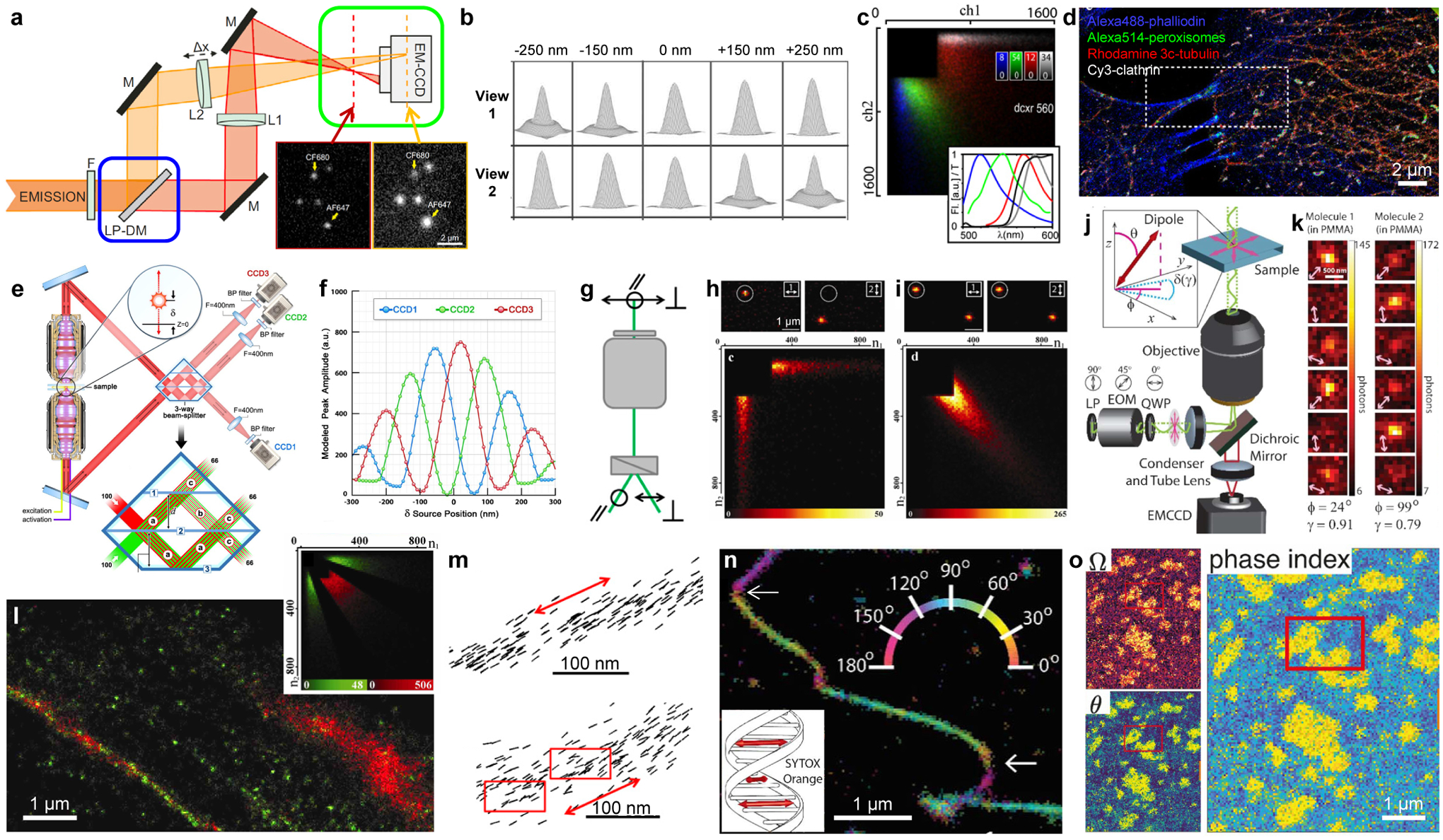
Figure 2.

Multi-view referencing for 3D, multicolor, and polarization SMLM. (a) Integration of biplane 3D imaging (green box: shifted focal planes between two views) and ratiometric color detection (blue box: fluorescence split into long- and short-wavelength components). LP-DM: long-pass dichroic mirror. Inset: single-molecule images obtained in the two views. (b) Simulated images of a point source at different axial positions for two views with a 500-nm focal shift. (c) Density heatmaps of photon counts recorded in the long- and short-wavelength channels, for individual molecules of four different dyes with their emission spectra (colored curves) and the transmission of the dichroic mirror (black curve) shown in the inset. (d) Four-color SMLM of a fixed cell by separating the four probes based on (c). (e) 3D-SMLM based on multiphase interference between fluorescence collected from two opposing objective lenses. (f) Expected brightness detected by the three cameras in (e) for single molecules at different axial positions. (g) Splitting the fluorescence into two orthogonal polarizations. (h) (top) Fluorescence images of single pcRhB molecules in PMMA, recorded in two channels of orthogonal polarizations. (bottom) Density heatmaps of photon counts recorded in the two channels for different single molecules. (i) Same as (h), but for pcRhB in mowiol. (j) An electro-optic modulator (EOM) rotates the linear polarization direction of the excitation laser in consecutive frames. (k) Resultant images of two rhodamine 101 molecules in PMMA, showing dissimilar changes in brightness in consecutive frames. (l) Classification of single β-actin-tdEosFP molecules in the SMLM image into immobile (green) and mobile (red) fractions based on the brightness in two channels of orthogonal polarizations (inset). (m) Single-molecule orientations measured for Alexa Fluor 488-phalloidin labeled to two actin fibers in fixed cells. Red arrows: averaged fiber direction. Red boxes: regions of structural heterogeneity. (n) Color-coded orientation-resolved SMLM image for SYTOX Orange labeled to a DNA strand in vitro. Arrows: abrupt bends. Inset: absorption dipole moment of the dye is perpendicular to the DNA axis. (o) Orientation-resolved SMLM image of Nile Red in a phase-separated supported lipid bilayer, shown as maps of solid angle (Ω), polar angle (θ), and combined phase index. (a) is from ref42. (b) is from ref31. (c,d) are from ref37. (e,f) are from ref33. (g,m) are from ref48. (h,i,l) are from ref47. (j,k,n) are from ref51. (o) is from ref49.
