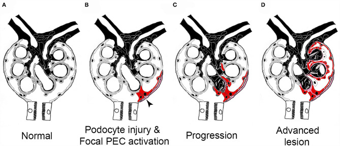
Figure 6.
Pathogenesis of sclerotic lesions in FSGS. (A) Normal glomerulus. gray, podocytes; white, parietal epithelial cells. (B) Podocyte injury or loss triggers focal PEC activation (red), PECs form an adhesion to the capillary tuft (arrow). (C) PECs take over parts of the capillary segment, capillaries are lost. (D) Advanced stage. Figure taken from (56) with modifications.

