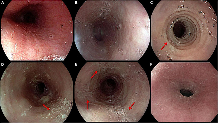
FIGURE 1.

Main endoscopic features of eosinophilic esophagitis. From top left: (A) Normal appearance of esophageal mucosa; (B) Edema. Pale mucosa with attenuation of the normal vascular pattern; (C) Rings. Trachealized esophagus with multiple concentric rings (arrow); (D) Exudates. Whitish small plaques not washable through water jet (arrow); (E) Furrows. Typical longitudinal furrows (arrows); (F) Stricture. Narrowing of esophageal lumen not passable by a standard scope (diameter around 9 mm).
