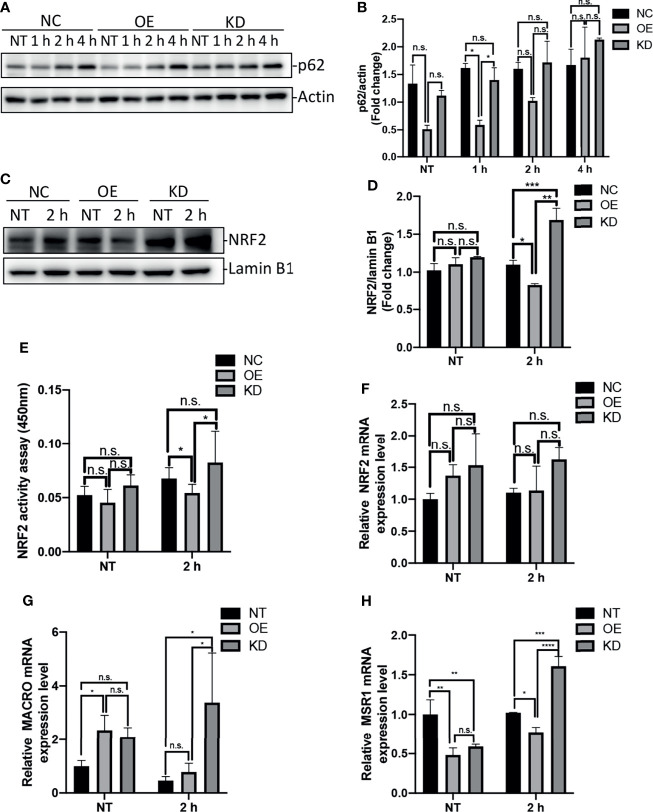
Figure 4.
Egr-1 suppressed NRF2 activation through reducing the levels of p62 in macrophages in response to P. aeruginosa infection. Egr-1 NC, OE and KD RAW264.7 cells were infected with P. aeruginosa PAO1 at an MOI of 10 for 1 h, 2 h, 4 h or left untreated (NT). Cell lysates were subjected to Western blotting for determining the p62 protein expression levels, and actin was used as a loading control. Blots are representative of three independent experiments (A). Densitometry analysis of the p62 protein levels was normalized to actin, and data were presented as fold change (B) (n = 3 ± SEM; n.s., not significant, *p < 0.05). The nuclear proteins were extracted from the Egr-1 NC, OE and KD cells infected with P. aeruginosa PAO1 for 2 h or left untreated, and subjected to Western blot analysis for examining the protein levels of NRF2 in nucleus. Blots are representative of three independent experiments (C). Densitometry analysis of the NRF2 protein levels in nucleus was normalized to lamin B1, and data were presented as fold change (D) (n = 3 ± SEM; n.s., not significant, *p < 0.05, **p < 0.01, ***p < 0.001). The nuclear extracts from NT and P. aeruginosa 2 h infected Egr-1 NC, OE and KD cells were subjected to transcription factor ELISA for determining NRF2 activity (E) (n = 6 ± SEM n.s., not significant, *p < 0.05). The total RNA isolated from NT and P. aeruginosa 2 h infected Egr-1 NC, OE and KD cells were reverse transcribed to cDNA and subjected to real-time quantitative PCR for analyzing the mRNA transcription levels of NRF2, MACRO and MSR1. The mRNA levels of NRF2, MACRO and MSR1 were normalized to endogenous control β-actin (F–H) (n = 3 ± SEM; n.s., not significant, *p < 0.05, **p < 0.01, ***p < 0.001, ****p < 0.0001).

