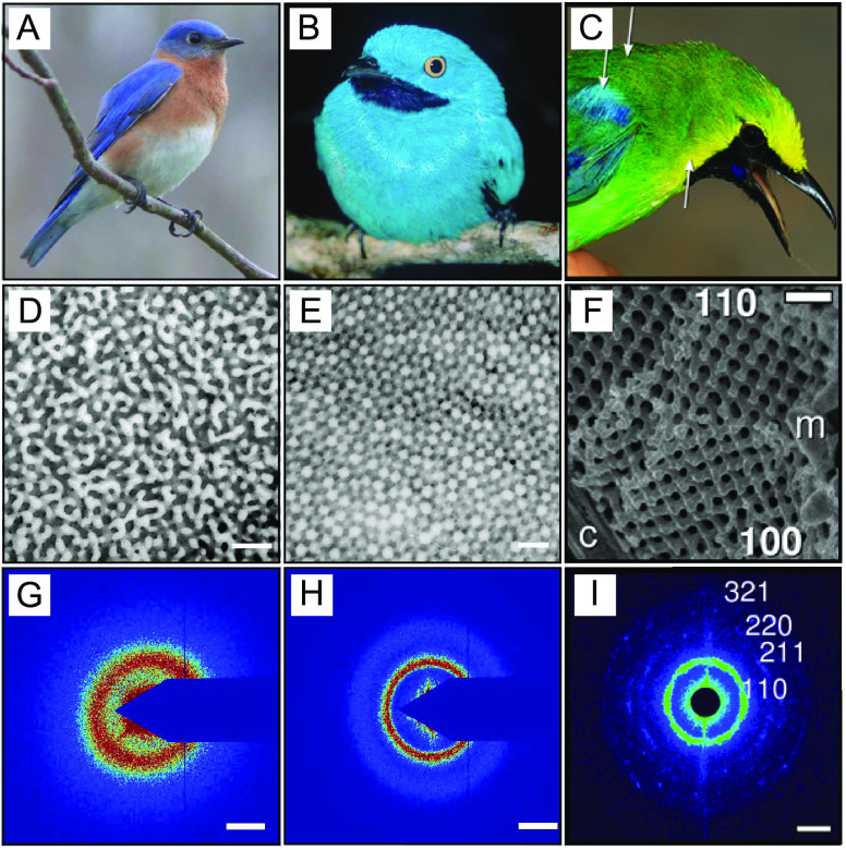
Figure 2.
Examples of coloration in bird feathers by structures formed during arrested phase separation. (A–C) Pictures of (A) Sialia sialis, (B) Cotinga maynana, and (C) C. cochinchinensis kinneari. (D–F) Scanning electron microscopy images of the underlying structure: (D) channel type, (E) sphere-type and (F) gyroid-type β-keratin and air nanostructure (G–I) X-ray scattering plots. Scale bars are (D–F) 500 nm and (G–I) 0.025 nm–1. Reproduced with permission from ref (40), Copyright 2009 Royal Society of Chemistry, and from ref (45), Copyright 2021 National Academy of Sciences.

