Figure 1.
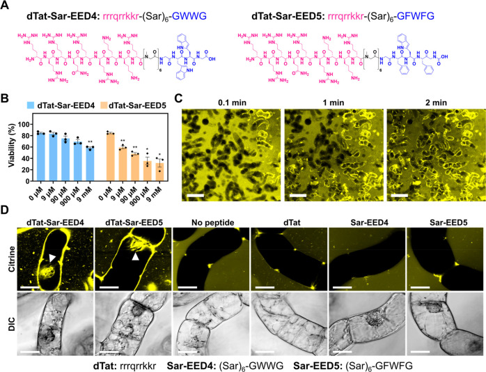
dTat-Sar-EED peptides mediate Citrine transduction into BY-2 cells. (A) Chemical structures of dTat-Sar-EED4 and dTat-Sar-EED5. (B) Viability of BY-2 cells determined by Evans blue assay after treatment for 1 h with various concentrations of either dTat-Sar-EED4 or dTat-Sar-EED5. Data from three biological replicates are represented as the mean ± standard error values. Statistical significance compared to the control (0 μM): *P < 0.05, **P < 0.01 based on Dunnett’s T3 test (n = 3). (C) Time-lapse confocal images of Citrine internalization into BY-2 cells treated with dTat-Sar-EED4 (90 μM). Scale bars, 2 mm. (D) Confocal images showing Citrine internalization into BY-2 cells treated for 1 h with Citrine alone (100 μg/mL, No peptide) or in combination with 90 μM dTat-Sar-EED4, dTat-Sar-EED5, dTat, Sar-EED4 or Sar-EED5. Differential interference contrast (DIC) images are shown below, with the amino acid sequence of each peptide below the images. White arrowheads indicate Citrine located in the nucleus. Scale bars, 20 μm.

