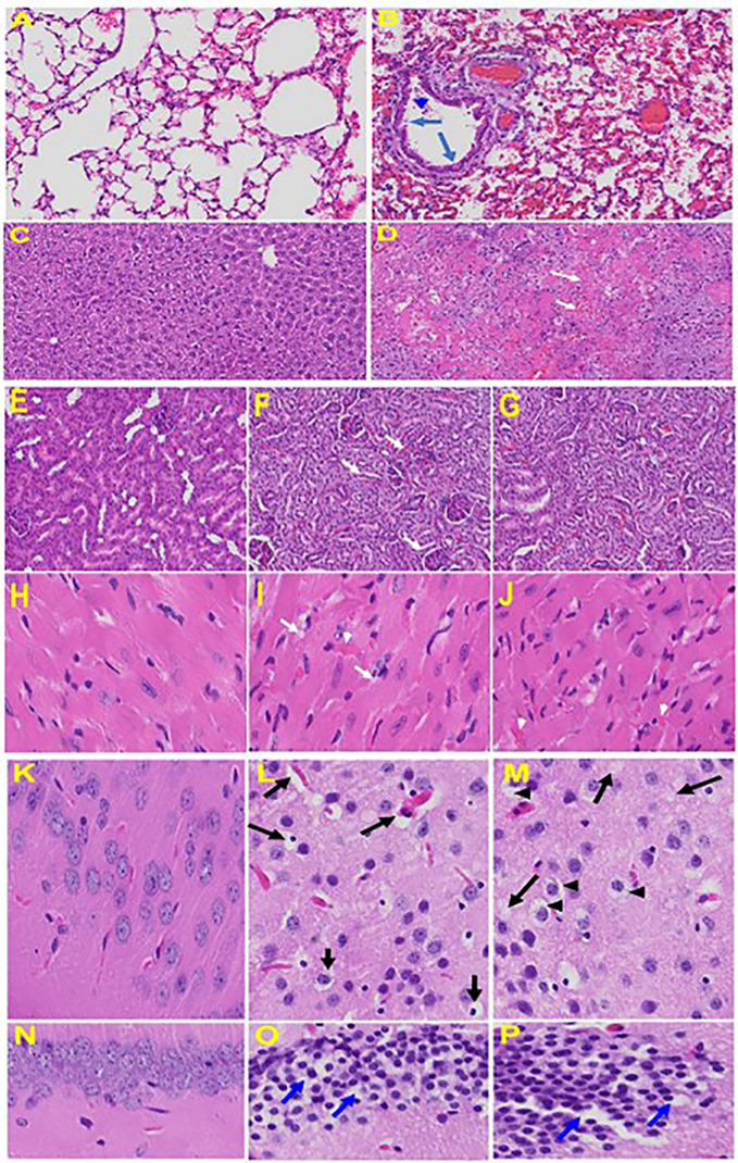
Figure 6.
Organs histology in infected and uninfected mice: Representative histological images of haematoxylin and eosin (H&E) stained lung tissue sections of normal mouse (A) and infected mouse lung (B). MHV-1-infected mice lung shows arterial endothelial swelling (hypertrophy, long arrow), inflammation and granular degeneration of cells (short arrow) and migration of leukocytes (arrowhead) into lung. Peribronchiolar interstitial infiltration, bronchiole epithelial cell necrosis and necrotic cell debris within alveolar lumens, alveolar exudation, infiltration, hyaline membrane formation and alveolar hemorrhage with red blood cells within the alveolar space and interstitial edema were observed in MHV-1-infected mice. Liver, kidney, heart, and brain tissue degenerative changes were observed in MHV-1-infected mice. MHV-1-infected mice at day 6 showed liver hepatocytes degeneration, severe cells necrosis [(D), long arrows] and hemorrhagic changes (short arrows) when compared to uninfected mice (C); Uninfected kidney (E) and tubular epithelial cells degenerative changes and vacuolation, and peritubular vessels congestion (F, G); Severe interstitial edema (arrows), vascular congestion and dilation (arrow heads) and red blood cells infiltrating between degenerative myocardial fibers were seen in the MHV-1 infected mice heart (I, J), as compared to uninfected mice (H). Vacuolation of cerebral matrix [arrows, (L, M)] and cytotoxic edema [arrow heads, (L, M)], congested blood vessels with perivascular edema [long arrows, (L, M)], as well as cytotoxic edema (short arrows) were seen in MHV-1infected mice brain cortex, and in hippocampus (O&P). Reproduced with permission from Paidas et al, Viruses; Published by MDPI, 2021.

