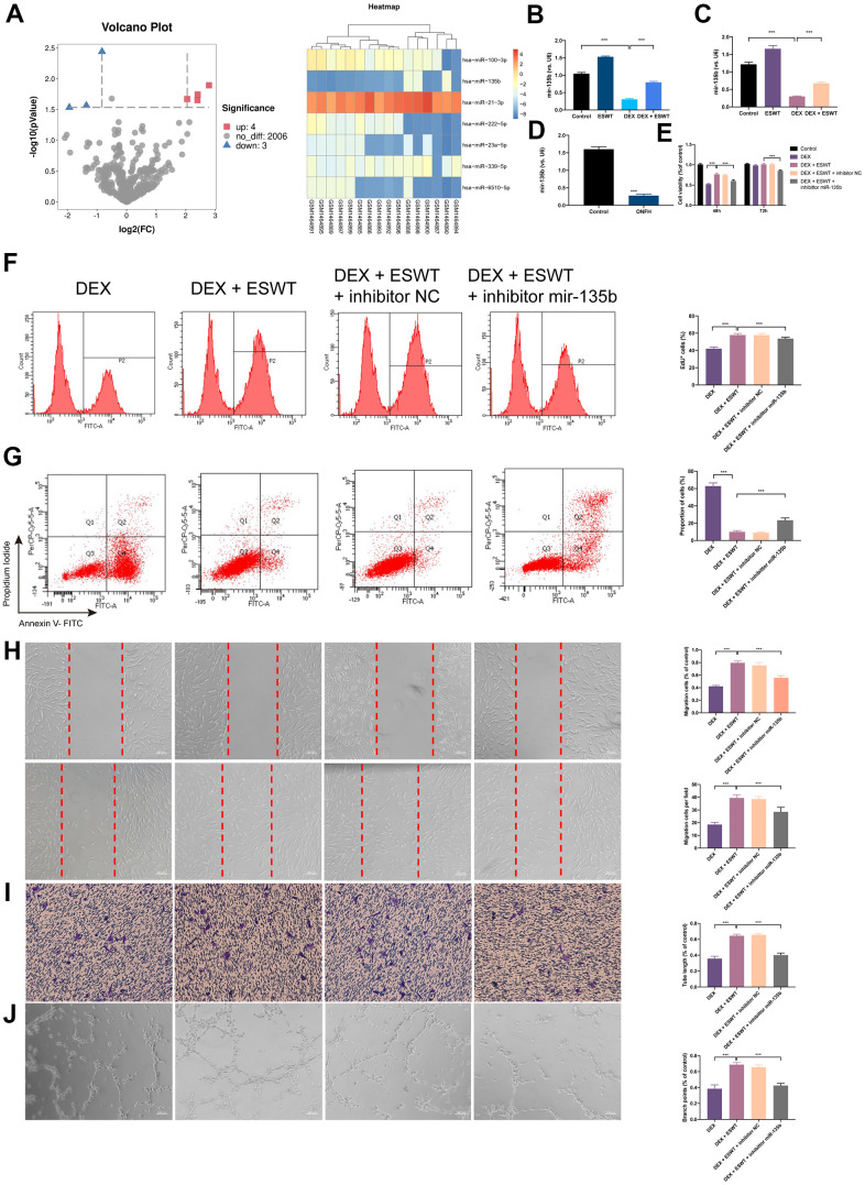
Figure 3.
Effect of miR-135b on endothelial cells treated with GCs. (A) Volcano plot and heat map of potential differentially abundance microRNAs; (B) expression of miR-135b in HUVECs; (C) expression of miR-135b in BMECs; (D) expression of miR-135b in femoral head tissue; (E) After transfection of inhibitor mir-135b, ECs were subjected to ESWT with 0.05 mJ/mm2, 1000 shots followed by DEX with 180 μM. cell viability examined by CCK-8 analysis in BMECs; (F) cell proliferation confirmed by EdU assay in BMECs; After transfection of inhibitor mir-135b, ECs were subjected to ESWT with 0.03 mJ/mm2, 1000 shots followed by DEX with 180 μM. (G) apoptosis rate of assessed through Annexin V-FITC/PI in BMECs; After transfection of inhibitor mir-135b, ECs were subjected to ESWT with 0.05 mJ/mm2, 1000 shots followed by DEX with 180 μM. (H) migration ability evaluated by wound healing assay in BMECs; (I) migration ability evaluated by Transwell assay in BMECs; (J) angiogenesis ability evaluated by tube formation assay in BMECs. n=3 **P < .01, ***P < .001.

