Figure 7.
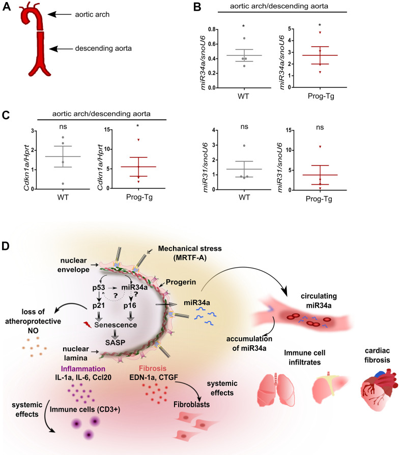
Analysis of circulatory SA-miRs at atheroprone aortic arch regions. (A) Schematic representation of aortic segment including aortic arch region and that of descending aorta. (B) Gene expression analysis of miR34a-5p (miR34a) and miR31-5p (miR31) and of (C) p21 (Cdkn1a) in WT and Prog-Tg aortic arch in comparison to descending aorta. n=4. Paired two-tailed Students t test, ns=non-significant *p<0.05. (D) Hypothetical model of intrinsic and extrinsic miR34-mediated senescence regulation in progerin-expressing endothelial cells. Mechanical stress particularly at vessel bifurcation leads to p53-linked senescence and miR34 upregulation. miR34 sustains senescence through positive feed-back mechanism acting on p53 but also separately by maintaining high levels of late senescence marker p16. Synergistic miR34-p53 action leads to elevation of SASP signaling and thus secretion of pro-inflammatory (Ccl20, IL-1a) and pro-fibrotic (CTGF, EDN-1a) factors with systemic effects on surrounding tissues leading to immune cell infiltrates and fibrosis in lung and liver and cardiac tissue. Systemic fibrosis and inflammation in tissues is further potentiated by increased release of miR34 in circulation.

