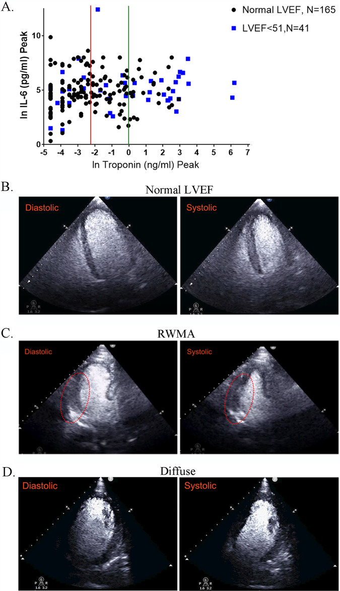
FIG 10.
Effect of SARS-CoV-2 infection on cardiac output in COVID-19 patients. (A) COVID-19 patients without prior cardiac disease were divided into two groups: patients with normal LVEF (n = 165) and those with a decreased LVEF of <51 (n = 41). We plotted the peak troponin I level (ng/mL) using a natural log scale (ln) and peak IL-6 (pg/mL). The red line indicates where troponin levels are at 0.1 ng/mL (ln = −2.3) and the green line indicates levels at 1 ng/mL (ln = 0). For the group with LVEF of <51, 20 out of 41 (50%) have troponin levels of >1 ng/mL, while the number with normal LVEF is 22/165 (13.3%). In another group of patients with LVEF of <51, 6/41 (14.6%) have troponin I levels greater than 20 ng/mL (ln = 3); none of the patients with normal LVEF have a troponin I level above 20 ng/mL. For troponin I, the difference is very significant by t test (P < 0.0001), when comparing the group with normal LVEF to the group with LVEF of <51. (B to D) Echocardiogram clips showing diastolic and systolic states. (B) “Normal” demonstrates an apical 4-chamber view (after administration of an ultrasonic enhancing agent) from a transthoracic echocardiogram obtained from a COVID-19 patient (see Movie S2 at https://iyengarlab.org). The findings are consistent with preserved left ventricular ejection fraction and no regional wall motion abnormalities (diastolic and systolic frames). The end-diastolic and end-systolic volumes are 104 mL and 35 mL, respectively, with a left ventricular ejection fraction of 66%. (C) Regional wall motion abnormality (RWMA) demonstrates an apical 3-chamber view (after administration of an ultrasonic enhancing agent) from a transthoracic echocardiogram obtained from a COVID-19 patient (see Movie S3 at https://iyengarlab.org). The findings are consistent with basal and mid infero-lateral wall hypokinesis, despite preserved left ventricular ejection fraction (diastolic and systolic frames). The RWMA is highlighted by the red oval. The end-diastolic and end-systolic volumes are 102 mL and 50 mL, respectively, with a left ventricular ejection fraction of 51%. (D) “Diffuse” demonstrates an apical 4-chamber view (after administration of an ultrasonic enhancing agent) from a transthoracic echocardiogram obtained from a COVID-19 patient (see Movie S4 at https://iyengarlab.org). The findings are consistent with diffuse left ventricular wall hypokinesis and mildly decreased left ventricular ejection fraction. The end-diastolic and end-systolic volumes are 160 mL and 90 mL, respectively, with a left ventricular ejection fraction of 44%.

