FIG 5.
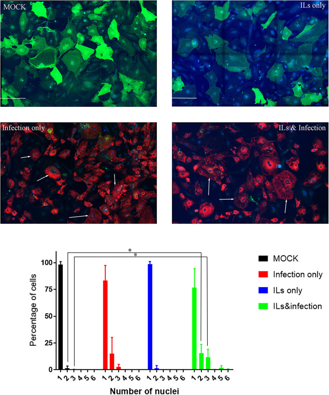
SARS-CoV-2 infection increases the percentage of multinucleated cells. Cells of CM line MSN-31-01S were treated with ILs and/or infected with SARS-CoV-2 at an MOI of 0.1 for 48 h. Shown is immunostaining of CMs for ACE2 (green), NP (red), and nuclear stain (blue). Mock-infected cells express ACE2 and predominantly have a single nucleus, but some double nuclei are detected. ILs did not alter the count of nuclei in these CM cells. Infection with SARS-CoV-2 at an MOI of 0.1 for 48 h does diminish the amount of ACE2 staining, and several cells are detected with 3 nuclei, as noted by the white arrows. Cells exposed to ILs and infection have an increase in the number of nuclei, where 3 to 5 nuclei are pointed out by the white arrows. The graph is the average number of nuclei counted between 3 independent experiments. Statistical significance was determined by unpaired t test, and a result was significant at P < 0.05 (*) comparing control to IL treatment and SARS-CoV-2 infection for 2 and 3 nuclei per cell. The scale bar in each image is 200 μm.

