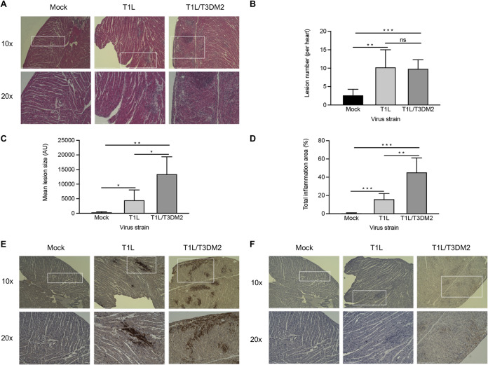
FIG 6.
T1L/T3DM2 is more myocarditic than T1L. Neonatal C57BL/6 mice were mock infected with PBS or inoculated orally with 104 PFU of T1L or T1L/T3DM2 viruses. At 8 days, the mice were euthanized, and the hearts were resected, paraffin embedded, and sectioned. Consecutive sections were stained with (A) H&E, (E) polyclonal reovirus antiserum, or (F) cleaved-caspase-3 antibodies. Sections were imaged at ×10 magnification (top) or 20× (bottom). Using H&E-stained sections, (B) cardiac lesion number, (C) lesion size (in arbitrary units [AU]), and (D) total area of inflammation (expressed as percentage of the total heart) were quantified (5 mice per group). Error bars indicate SD. *, P < 0.05; **, P < 0.01; ***, P < 0.001; ns, not significant (determined by Student's t test).

