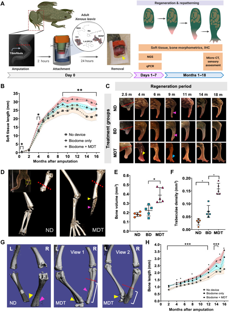
Fig. 1. Twenty-four–hour multidrug treatment delivered by bioreactor improves repatterning in regenerates.
(A) Xenopus hindlimbs were amputated and inserted into silk-based hydrogel devices (yellow arrow) for 24 hours. Hydrogels contained a multidrug treatment (MDT) or no added factors (BD). Some animals received no device (ND). (B) Regenerated MDT hindlimbs were longer than the BD and ND groups by 2.5 mpa, as indicated by growth beyond resection site (red dashed line). At 4 mpa, vascularized structures developed at the distal extension of MDT, but not BD or ND regenerates. At 9 mpa, digit-like projections appeared (blue arrow), contrasting the hypomorphic spikes of BD and ND regenerates (pink arrows). (C) Soft tissues of MDT animals were consistently longer than BD or ND from 8 mpa [F(2,19) = 61.9, P < 0.05]. (D) Micro-CT reconstructions revealed increases in regenerate bone length (yellow arrow) and volume, as well as diffuse bone (purple arrow). (E) Volumetric quantification confirmed increased growth in MDT regenerates [F(2,15) = 11.15, P < 0.001]. (F) Tuberculae complexity increased in MDT-exposed tissue. (G) Micro-CT reconstructions show reestablishment of fine bone structures in MDT regenerates (purple arrow). (H) Longitudinal x-ray imaging indicates gradual accelerated bone growth in MDT [F(2,19) = 31.04, P < 0.05]. Means and SD are presented. *P < 0.05; **P < 0.01; ***P < 0.001.

