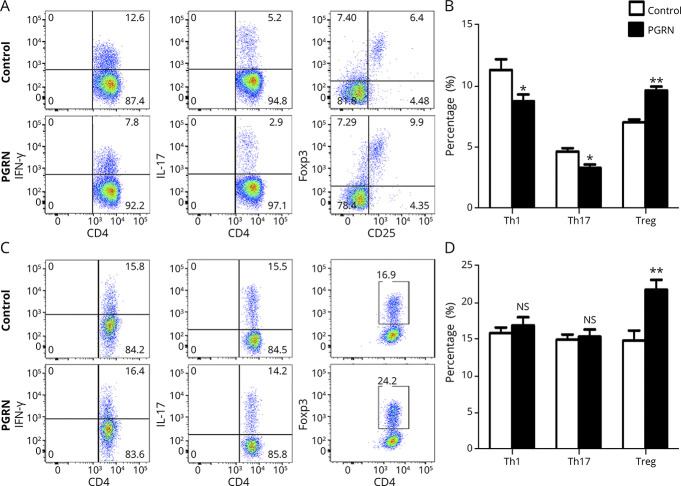
Figure 4. Recombinant PGRN Inhibits IRBP-Reactive Th1 and Th17 Cell Expansion and Promotes IRBP-Reactive Treg Cell Expansion.
(A and B) Spleen cells from EAU mice were stimulated with IRBP651–670 in the presence or absence of rPGRN for 72 hours and then assessed for intracellular expression of IFN-γ, IL-17, and Foxp3 by CD4+ T cells by flow cytometry (n = 8). (A) Representative flow cytometry dot plots. (B) Histograms of IFN-γ, IL-17, and Foxp3 by CD4+ T cells (Th1, Th17, and Treg, respectively). (C and D) Naive CD4+ T cells from spleen cells of normal C57BL/6 mice were stimulated with Th1, Th17, and Treg cell polarization conditions in the presence or absence PGRN for 72 hours and then assessed for intracellular expression of IFN-γ, IL-17, and Foxp3 by CD4+ T cells by flow cytometry (n = 8). (C) Representative flow cytometry dot plots of the 2 groups. (D) Histograms of Th1, Th17, and Treg of the 2 groups. Data are shown as mean ± SEM from 2 independent experiments. *p < 0.05, **p < 0.01, and NS, not significant. EAU = experimental autoimmune uveitis; IL = interleukin; IFN = interferon; PGRN = progranulin; RT-PCR = real-time PCR; Th = T helper; Treg = regulatory T.

