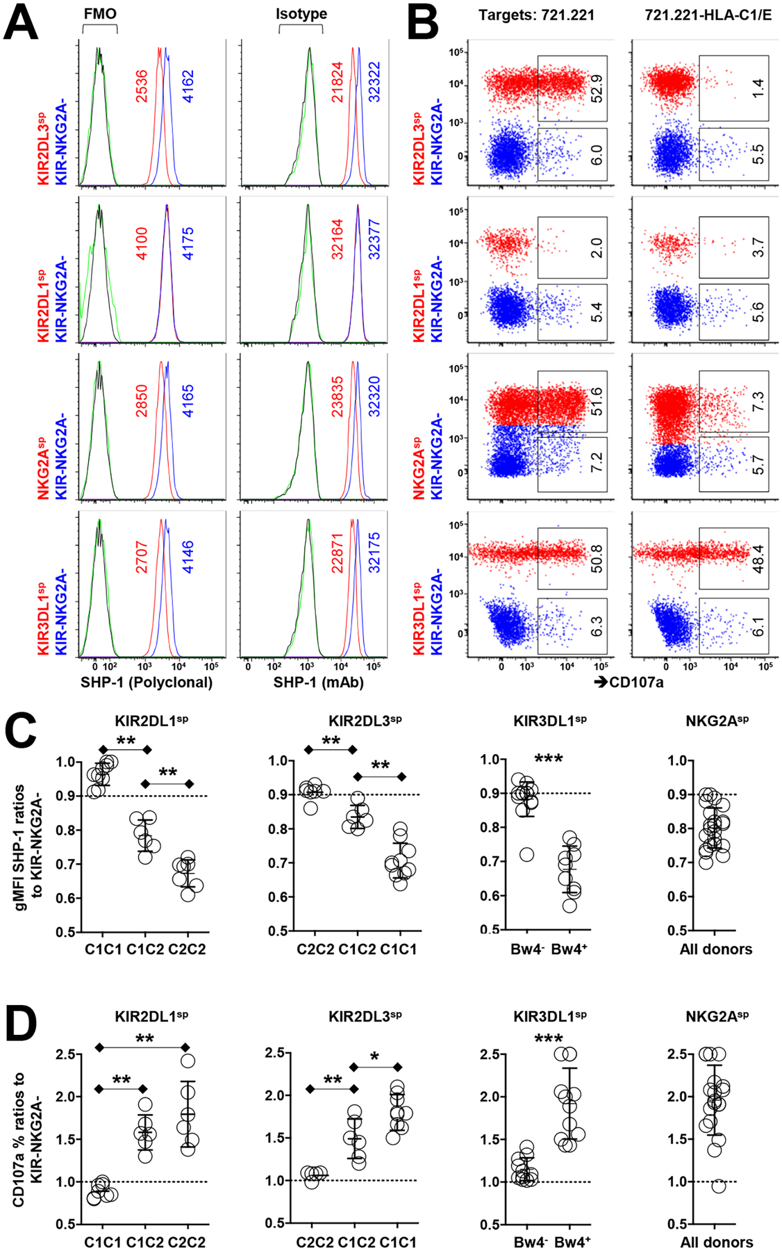
Fig. 1. Low SHP-1 abundance in human NK cells correlates with high responsiveness.

(A) Flow cytometric analysis of relative SHP-1 abundance among the indicated human NK cell subsets (stained with the polyclonal antibody C19 or the monoclonal antibody Y476) from an HLA-C1+C2−Bw4+ donor. gMFI values of SHP-1 are indicated in the plots. Staining controls are indicated in green for KIR+ or NKG2A+ cells and in black for KIR−NKG2A− cells. FMO, fluorescence minus one. (B) Analysis of the degranulation of the indicated NK cell subsets in response to treatment with 721.221 cells or HLA-C1/E-expressing 721.221 cells. Red populations are KIR-expressing or NKG2A-expressing cells. The indicated percentages of positive cells in the dot plots were determined as a percentage of the KIR+ and NKG2A+ single-positive (sp) cells or the KIR−NKG2A− NK cells. (C) Ratio of relative SHP-1 abundance (as determined with the Y476 antibody) among cells single-positive for KIR or NKG2A from 20 KIR haplotype-A homozygous donors. (D) Ratio of the degranulation activity of the indicated NK cell subsets to K562 cells from the indicated HLA genotypes. Data are representative of three (A and B) independent experiments. Data are means ± SD in (C) and (D), n = 20 human donors. *P < 0.05; **P < 0.01; ***P < 0.001; Friedman test and post hoc pairwise Wilcoxon test for multi-group comparison were used. Each symbol represents an individual human donor.
