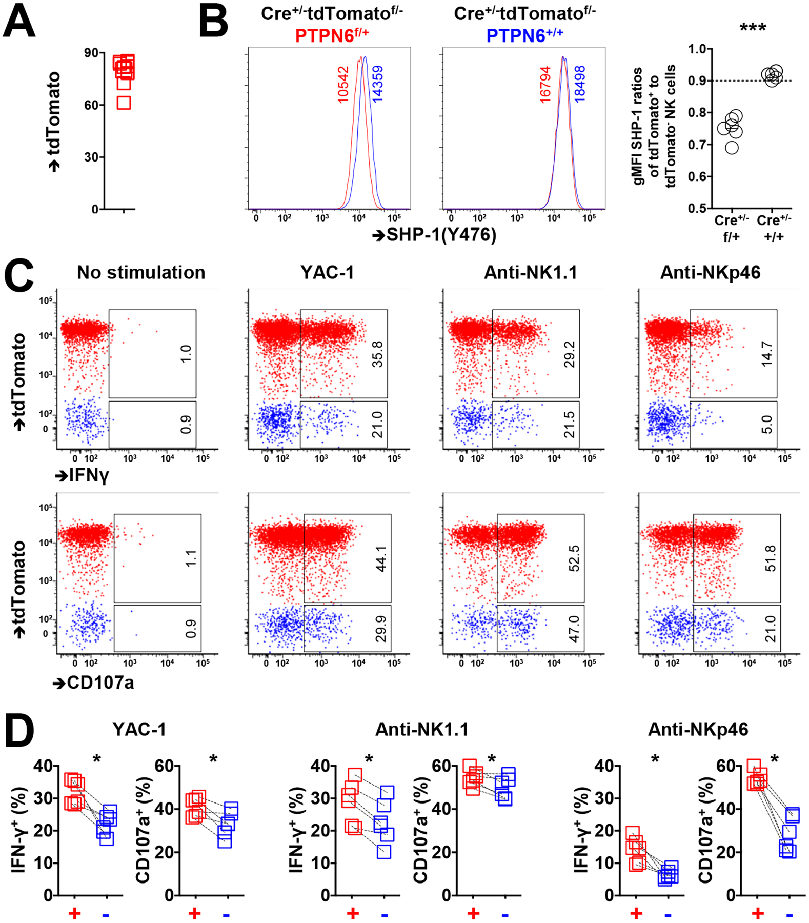
Fig. 4. NK cells have enhanced activity when SHP-1 is reduced.

(A) TdTomato abundance among NKp46+ splenic NK cells from CreERT2+/−Ptpn6fl/+tdtomatofl/− mice 3 days after tamoxifen administration. (B) SHP-1 abundance among tdTomato+ and tdTomato− NK cells from CreERT2+/−Ptpn6fl/+tdtomatofl/− and CreERT2+/−Ptpn6+/+tdtomatofl/− mice 3 days after tamoxifen administration. (C and D) IFN-γ production and CD107a activity among tdTomato+ and tdTomato− NK cells in response to YAC-1 cells or treatment with crosslinking anti-NK1.1 or anti-NKp46 antibodies. Paired symbols indicate populations from the same mouse. Results are representative of three independent experiments. The indicated percentages of positive cells in the dot plots were determined as percentages of the tdTomato+ and tdTomato− NK cells. Data are means ± SD in (B), n = 11 mice. *P < 0.05; ***P < 0.001; Mann-Whitney U test (B), and Nonparametric Wilcoxon signed rank sum test (D) were used. Each symbol represents an individual mouse.
