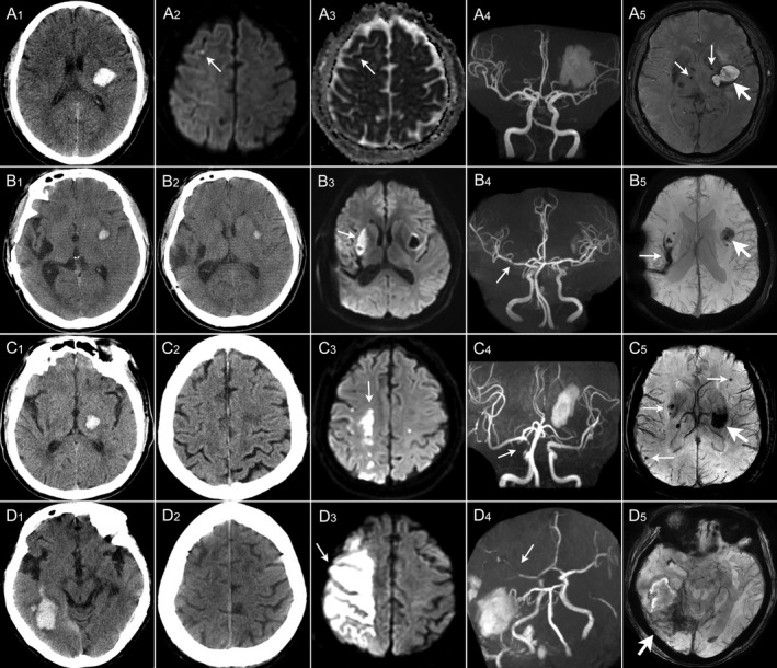
Figure 2.

Imaging examples of large and small DWI lesions in four patients with acute intracerebral hemorrhage. (A) In a 69‐year‐old man with left BG hemorrhage (A1), diffusion‐weighted imaging (DWI) sequence (A2) shows a small DWI hyperintensity lesion (thin arrow) in the right frontal lobe with corresponding low signal intensity on ADC sequence (A3) and magnetic resonance angiography (MRA) (A4) shows no stenosis in the intracranial artery; susceptibility‐weighted imaging (SWI) (A5) shows a hematoma (thick arrow) in left BG and CMBs in bilateral BG (thin arrow). (B) In a 43‐year‐old man with left putamen hemorrhage (B1), DWI sequence (B3) shows a large DWI hyperintensity lesion (thin arrow) in the deep branch territory of right middle cerebral artery (MCA) (right BG) which was normal on CT (B2) at admission and MRA (B4) shows mild stenosis (thin arrow) in M1 segment of right MCA; SWI (B5) shows a hematoma (thick arrow) in left putamen and an old hemorrhagic cavity in right BG (thin arrow). (C) In a 70‐year‐old man with left thalamus hemorrhage (C1), DWI sequence (C3) shows borderzone infarct (thin arrow) in borderzone territories of MCA and ACA which was normal on CT (C2) at admission and MRA (C4) shows moderate stenosis in the terminal of right internal carotid artery (thin arrow); SWI (C5) shows a hematoma in left BG (thick arrow) and multiple CMBs (thin arrows). (D) In a 68‐year‐old woman with right temporal lobe hemorrhage with subarachnoid extension (D1). DWI sequence (D3) shows a large DWI hyperintensity lesion (thin arrow) in right MCA territories (right frontal and parietal lobes) which was normal on CT (D2) at admission and MRA (D4) shows severe stenosis to occlusion in M2 segment of right MCA (thin arrow); SWI (D5) shows a hematoma in right temporal and occipital lobes (thick arrow).
