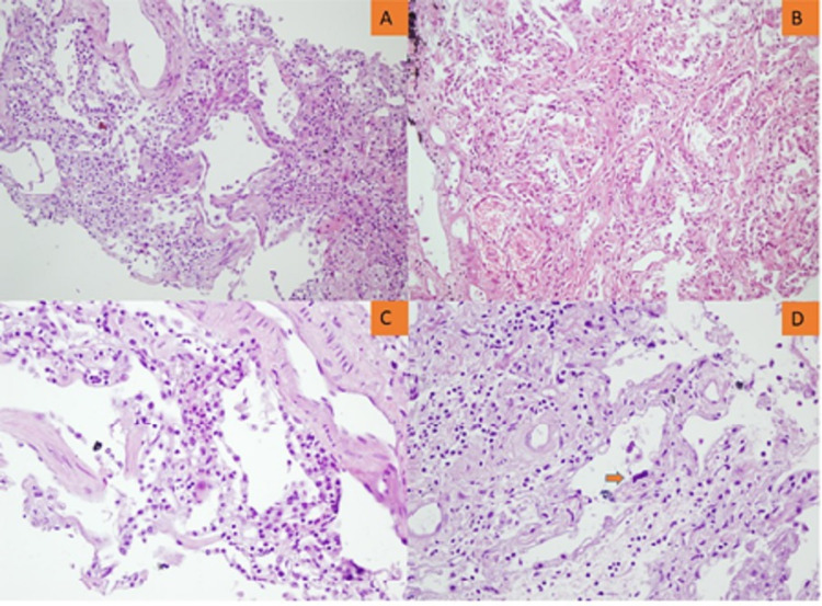
Figure 3. Lung microscopy showing the late organizing phase of ARDS.
A. Septal thickening with interstitial cellular increase (x100). B. Septal thickening with interstitial cellular and collagenous increase (x200). C. Interstitial inflammatory cellular infiltration (x400). D. Interstitial thickening with cellular and collagenous increase, atypical type II pneumocyte (orange arrow).
ARDS: acute respiratory distress syndrome.

