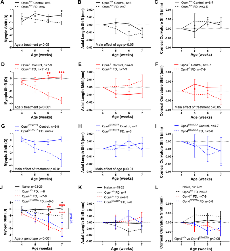
Figure 2.
Larger response to form-deprivation (FD) in melanopsin deficient mice. A) FD caused a myopic shift in Opn4+/+ (solid line) mice after 3 weeks (dashed line, p<0.05). B) There was no effect of FD on axial length in Opn4+/+ mice, but the axial length shift changed with age (p<0.05) C). Corneal curvature did not significantly change with FD. D-F) Same as in A-B but for Opn4−/− mice. There was a significant myopic shift with FD (p<0.001) but no change in axial length. However, cornea radii became shorter in the FD eye (p<0.05), developing a steeper corneal curvature. G-I) Same as in A-B but for Opn4DTA/DTA mice. FD led to a significant myopic shift (p<0.01) but no change in axial length or corneal curvature. J) Myopic shifts compared between naïve (solid black), Opn4+/+ FD (dashed black), Opn4−/− FD (dashed red), and Opn4DTA/DTA FD (dashed blue) mice. Opn4+/+ (black), Opn4−/− (red), and Opn4DTA/DTA (blue) mice. Opn4−/− mice show significantly greater susceptibility to FD than Opn4+/+ mice (p<0.001) and both Opn4−/− and Opn4DTA/DTA mice responded similarly to FD (p=0.34). Black and red asterisks denote significance between naïve compared to Opn4+/+ and Opn4−/− mice, respectively. K) There was no difference in axial length shift across genotypes. L) Corneal curvature after FD was only significantly different between Opn4+/+ and Opn4DTA/DTA mice (p=0.03). Axial length and corneal curvature values were normalized to 4 weeks of age. Data are presented as mean ± SEM. Comparisons between treatment (FD vs control) and genotypes were performed with MEA (or ANOVA) with HSK where *p<0.05, **p<0.01, and ***p<0.001.

