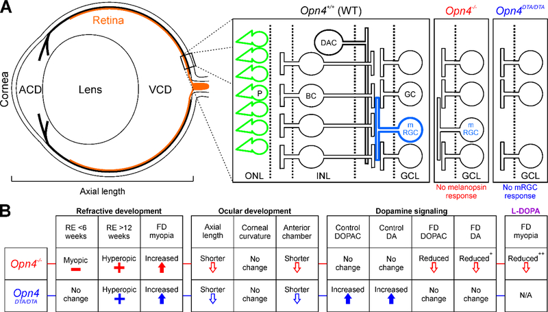
Figure 5.
Major development and signaling changes in the absence of melanopsin. A) Schematic of the mouse eye showing the major ocular development parameters (left, ACD = anterior chamber depth, VCD = vitreous chamber depth) and the major retinal pathways associated with melanopsin-mediated signaling (right). In Opn4+/+ mice, both photoreceptors (P, green) and mRGCs (light blue) directly detect light. mRGCs also receive photoreceptor-mediated synaptic input through bipolar cells (BC) and have interactions with dopaminergic pathways (DAC). In Opn4−/− mice (red), mRGCs still respond to light through photoreceptor-mediated synaptic signaling but no longer have intrinsic melanopsin light responses. In Opn4DTA/DTA mice (blue), mRGCs are not present and the retina loses both intrinsic mRGC and synaptic melanopsin responses. B) Summary table of the effects of melanopsin signaling disruption on refractive development, ocular development, and dopamine signaling. RE = refractive error. FD = form-deprivation. DA = dopamine. All parameters are in comparison to Opn4+/+ or the un-treated condition. + indicates p<0.10. ++ indicates p<0.10 in comparison to Opn4−/− mice without L-DOPA treatment.

