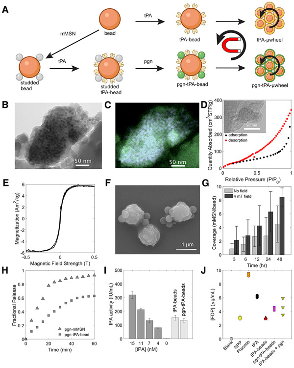
Fig. 3:
Synthesis and characterization of magnetic mesoporous silica nanoparticles (mMSN) and their coupling to superparamagnetic beads. A) Schematic of tPA-functionalization, mMSN-bead coupling, and plasminogen (pgn) loading to create tPA- and tPA-pgn-μwheels. B) Transmission electron microraph (TEM) of mMSN. Darker areas are Fe3O4 domains incorporated into the silica matrix. C) High-angle annular dark field energy-dispersive X-ray (HAADF-EDS) spectrum showing iron oxide domains (blue) distributed throughout silica matrix (green). D) Representative BJH isotherm data with TEM image of pore structure (inset). E) Magnetization profile for mMSN. F) Scanning electron micrograph (SEM) of studded beads. G) Coverage of mMSN to beads as a function of mixing time in the with and without a 4 mT magnetic field. H) Plasminogen release profile for pgn-mMSN and pgn-tPA-beads at a number density of 106/μL. Fractional release is normalized to the total plasminogen loading as measured by spectrometry. I) Activity of 105/μL bead populations compared to solvated tPA, measured using a fluorogenic substrate. J) Concentration of FDP in NPP after 60 min incubation with bead populations having an equivalent activity to 50 nM tPA.

