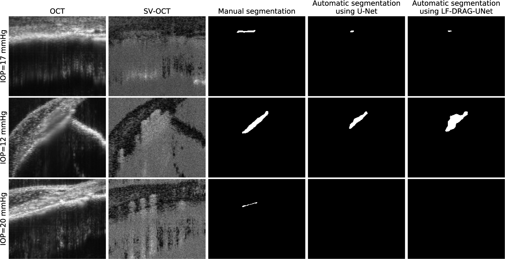
Figure 8.

Limitations of CNNs for automatic SC segmentation. Columns from left to right represent the intensity OCT image, SV-OCT image, ground truth obtained by manual segmentation, segmentation obtained by U-Net, and segmentation obtained by the proposed LF-DRAG-UNet model. Low contrast, shadowing due to superficial blood vessels, and small SC size can lead to incorrect segmentations.
