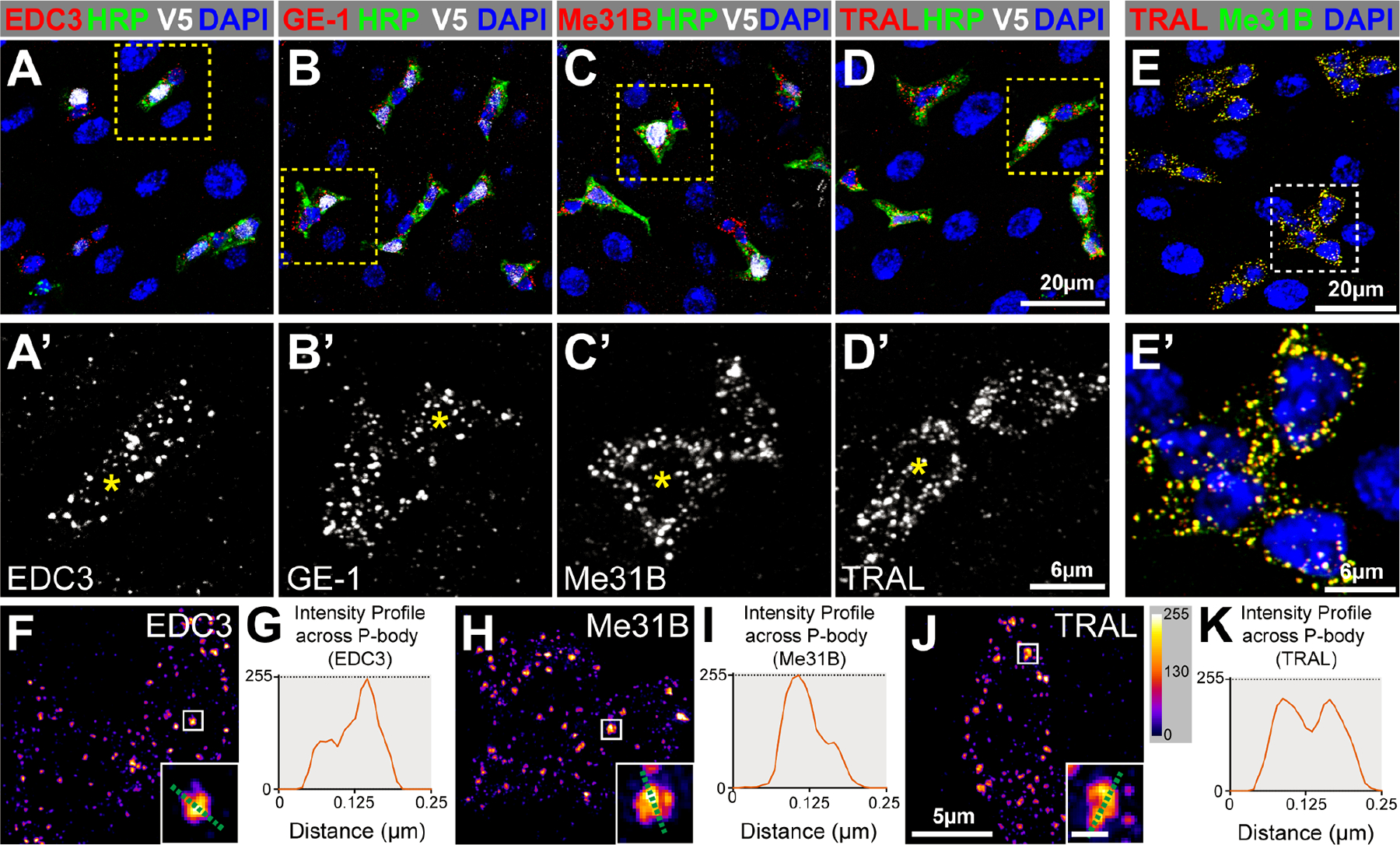
Figure 1. Intestinal progenitor cells contain P-bodies.

(A-D) Adult posterior midguts (PMGs) stained for EBs (V5, white), progenitor cells (HRP, green), all cells (DAPI, blue) and either EDC3, GE-1, Me31B, or TRAL (red). A’-D’ are enlargements with EBs labeled (asterisks). (E) PMG stained for TRAL (red), Me31B (green), and DAPI (blue). (F, H, J) Super-resolution micrographs of progenitor cells stained for EDC3, Me31B, or TRAL pseudocolored using intensity scale in J; insets are from boxed regions (scale 0.125μm). (G, I, K) Pixel-by-pixel intensity profiles along the green dotted line shown in insets. Full genotypes listed in Data S1F. See also Figure S1.
