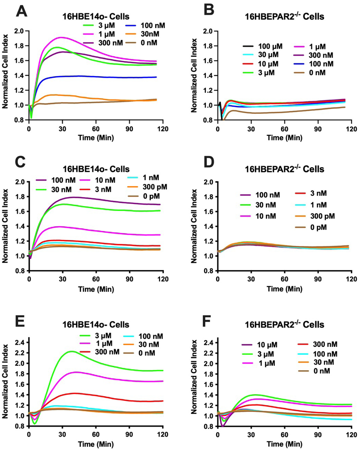
Figure 2: The in vitro physiological responses of human bronchial epithelial cells following addition of select PAR2 activators.

Each panel (A - F) represents the impedance responses (Cell Index) measured each minute following the addition of PAR2 agonist. Agonist concentrations were chosen to reflect full responses in 16HBE14o− cells and reduced in ½ log steps Traces represent the average of four experiments. Concentration-dependent response to 2at-LIGRL-NH2 for 16HBE14o− (A) or 16HBEPAR2−/− (B) cells. Concentration-dependent response to trypsin for 16HBE14o− (C) 16HBEPAR2−/− (D) cells. Concentration-dependent response to elastase for 16HBE14o− (E) 16HBEPAR2−/− (F) cells. The lack (2at-LIGRL-NH2; trypsin)- or severely reduced (elastase)-induced physiological responses in the 16HBEPAR2−/− cell line demonstrate a necessity for PAR2 expression for in vitro physiological response.
