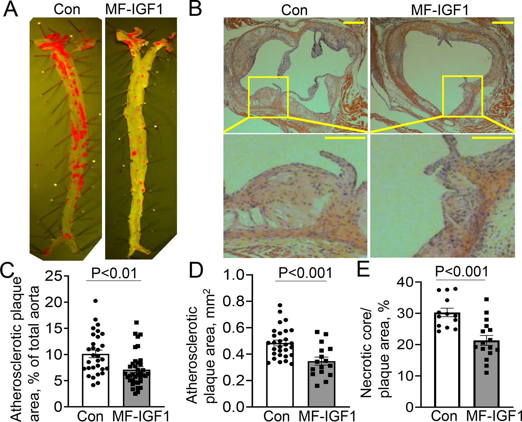
Figure 2: Macrophage-specific IGF-1 overexpression reduced atherosclerosis.

Macrophage-specific IGF-1 overexpressing (MF-IGF1 mice) and control mice (Con) were fed with a high-fat diet. A, C. Enface analysis of atherosclerotic burden (N=30–37 mice per group). B, D. H&E stained cross-sectional aortic valve sections to assess lesional area (N=16–29 mice per group). B Insert: magnified view of lesions showing that plaque in MF-IGF1 mice is less cellular. Scale bar, 100 μm. E. Necrotic core area in atherosclerotic plaque (N=14–15 mice per group). All statistical tests are Student’s two-tailed t-test.
