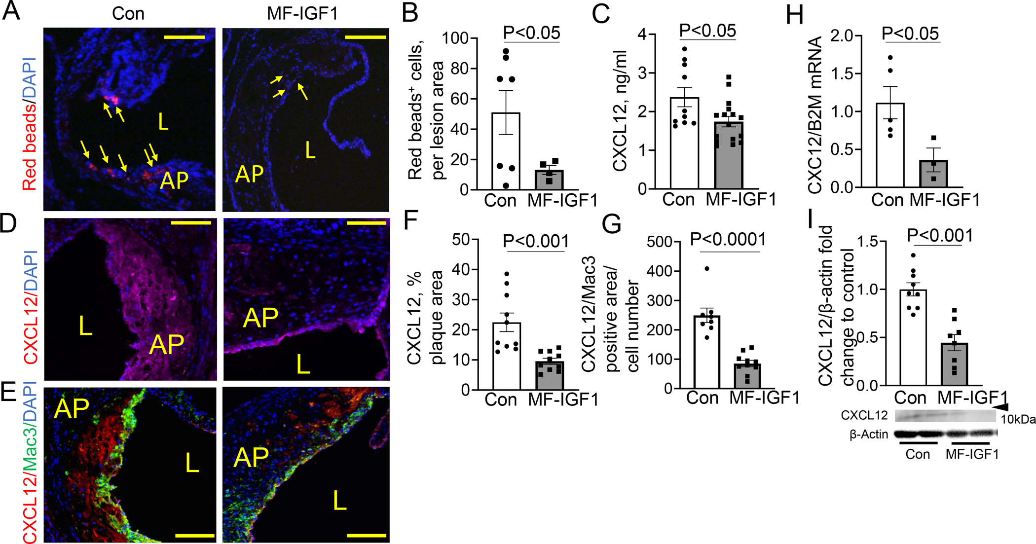
Figure 4: Macrophage-specific IGF-1 overexpression reduced monocyte recruitment into atherosclerotic plaque and decreased CXCL12 chemokine expression.

A, B. Monocyte recruitment was measured by quantification of plaque levels of red beads after normalization to plaque size. Arrows, plaque red spots (N=4–7 mice per group). C. Circulating levels of CXCL12 measured by ELISA (N=8–11 mice per group). D, F. CXCL12 positive area was normalized to plaque area (N=10 mice per group). E,G. CXCL12/Mac3 positive area was normalized to cell number (N=8–10 mice per group). H. CXCL12 mRNA levels in LCM plaque isolates (N=3–5 mice per group). I. CXCL12 levels in peritoneal macrophages were quantified by immunoblotting. Predicted weight 7–14kDa. (N=8–9 mice per group). Scale bar, 100 μm. All statistical tests are Student’s two-tailed t-test, except B, which had a Welch’s correction due to differences in SDs. AP=Atherosclerotic Plaque, L=lumen
