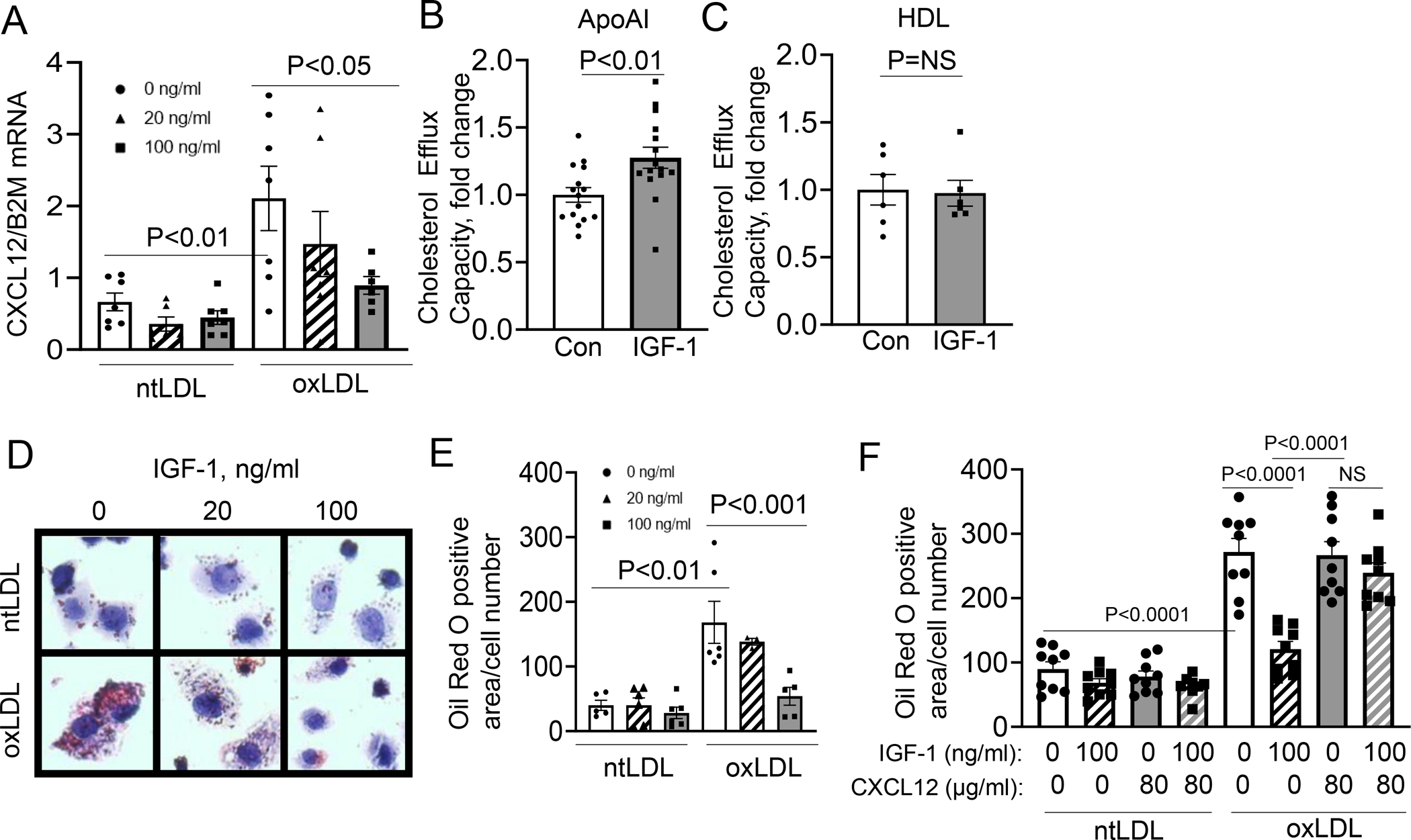
Figure 6: IGF-1 reduced formation of THP-1 macrophages-derived foam cell formation.

THP-1-derived macrophages were pretreated with IGF-1 and then treated with oxLDL or ntLDL. A. CXCL12 mRNA levels in THP-1 macrophages (N=3 wells per group per experiment, 3 independent experiments). B.10μg/ml ApoA1 was used as a cholesterol acceptor and cells were pretreated with IGF-1 then treated with oxLDL. Cholesterol efflux capacity was normalized to 0ng/ml IGF-1 treatment (Con). (N=4–7 wells per group per experiment, 3 independent experiments). C. 200μg/ml HDL was used as a cholesterol acceptor and cells were pretreated with IGF-1 then treated with oxLDL. Cholesterol efflux capacity was normalized to 0ng/ml IGF-1 treatment (Con). (N=3 wells per group per experiment, 2 independent experiments). D. Representative images of Oil Red O-stained macrophages. E. Quantitative data. (N=3 wells per group per experiment, 3 independent experiments). F. Cells were treated with IGF-1 and/or CXCL12 and then Oil Red O staining was used to quantify neutral lipids. (N=3 wells per group, 3 independent experiments). All statistics are one-way ANOVA except in D and E, which used a Student’s two-tail t-test. B used a Tukey’s post-hoc test, and C used Dunnett’s post -hoc test.
