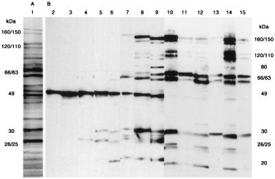
FIG. 2.
(A) Lane 1, protein profile of whole-cell sonicates of H. pylori ATCC 43504. Proteins were separated on an SDS–8 to 16% PAGE gradient gel and stained with Coomassie brilliant blue R-250. (B) The time course of Western blotting patterns with sera from H. pylori-inoculated gerbils. Lane 2, serum from a noninoculated gerbil; lanes 3 to 15, sera from inoculated gerbils. Times after inoculation were as follows: lanes 3 and 4, 2 weeks; lanes 5 and 6, 4 weeks; lanes 7 and 8, 8 weeks; lane 9, 12 weeks; lanes 10 and 11, 26 weeks; lanes 12 and 13, 38 weeks; lanes 14 and 15, 52 weeks. Lanes 10, 12, and 14 show sera from the ulcer group; lanes 11, 13, and 15 show sera from the hyperplastic group. Sera were diluted as follows: lanes 2 to 6, 1:100; lanes 7 to 9, 1:1,000; lanes 10 to 15, 1:4,000. Molecular masses are indicated in kilodaltons.

