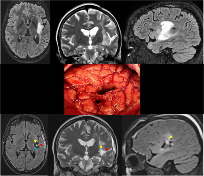
Figure 3.
Patient 5. Upper: Preoperative axial FLAIR (left), coronal T2 (middle), and sagittal FLAIR-weighted MRI (right) revealing a left posterosuperior insular LGG in a 36-year-old woman who experienced seizures. Middle: Intraoperative view (the anterior part of the left hemisphere is on the right and its posterior part is on the left) after resection in awake patient, achieved up to eloquent structures, both at cortical and subcortical levels. Number tags show positive DES sites, i.e., ventral premotor cortex eliciting anarthria when stimulated (1, 2), and primary somatosensory cortex of the face (3) within the retrocentral gyrus. According to this cortical mapping, a transopercular surgical approach has been selected via the alSMG (through the parietal operculum) and the superior temporal gyrus. In addition to the functional cortical areas, DES of white matter tracts allowed the detection of the critical subcortical neural networks (lSLF, AF, and somatosensory TCP). Lower: Postoperative axial FLAIR (left), coronal T2 (middle), and sagittal FLAIR-weighted MRI (right) demonstrating NTR. The neurological examination was normal 3 months following surgery. A diffuse WHO grade II astrocytoma was diagnosed, and no adjuvant treatment was administrated, with a regular surveillance. LGG, low-grade glioma; DES, direct electrical stimulation; alSMG, anterolateral part of the supramarginal gyrus; lSLF, lateral part of the superior longitudinal fasciculus (red circle); AF, arcuate fasciculus (yellow circle); TCP, thalamocortical pathway (blue circle); NTR, near total resection.

